The direct answer is that electron microscopy transforms nanoscale observations into actionable pilot-plant decisions. Techniques like TEM and SEM‑based EPMA quantify the crystallite size, dispersion, and spatial distribution of the active metal. This data directly determines the catalyst effectiveness factor, the pressure drop across the bed, the hot‑spot risk in exothermic reactions, and ultimately the reactor configuration, particle size, and operating space velocity you choose for your pilot campaign.
The core value of electron microscopy in pilot‑plant reactor design is its power to predict transport limitations before they appear at the pilot scale. By converting TEM‑derived dispersion and crystallite size into an internal effectiveness factor, and by using SEM‑mapped metal profiles to spot poisoning gradients, you can select a particle size, shape, and operating window that maximize kinetic data quality while avoiding fluid‑dynamic failures like channelling, excessive pressure drop, or fluidized‑bed collapse.
Decoding the Nanoscale with Electron Microscopy
What TEM and SEM Actually Quantify
TEM gives you the metal’s true “working surface”. It directly images crystallites in the range of 20–130 Å, delivering a dispersion value that tells you how many active atoms are exposed. This number is the anchor for all mass‑transfer calculations.
SEM‑based EPMA reveals how that active material is distributed inside a pellet. Cross‑sectional X‑ray maps highlight whether the metal forms an egg‑shell, egg‑yolk, or uniform profile. An uneven distribution at the micron scale signals preparation flaws or impurity gradients that will later show up as premature deactivation or hot spots in your pilot reactor.
Bridging from Micrographs to Reactor‑Scale Physics
The internal diffusion resistance inside catalyst pores is the first bridge. A high‑dispersion catalyst with small crystallites still relies on the pore network to bring reactants to those sites. The longer the diffusional path—determined by the pellet’s macroscopic particle size—the steeper the concentration drop.
That directly sets the internal effectiveness factor, η. TEM‑verified dispersion enables you to model how much of the pellet is actually participating. Once you know η, you can back‑calculate the true intrinsic kinetics from pilot‑plant data rather than mistaking a mass‑transfer limitation for slow chemistry.
How Microscopy‑Driven Data Shapes Reactor Design
Selecting Particle Size and Bed Geometry
For a fixed‑bed pilot plant, particle size is a balancing act. Smaller particles cut the diffusion path, pushing η toward 1.0 and giving you kinetic‑controlled data. But they also increase the bed pressure drop, which can crush catalysts, distort flow, and make data impossible to interpret.
Microscopy tells you where the cliff edge is. If TEM shows a very high crystallite dispersion, moving to a slightly larger particle may still keep η acceptable while dramatically lowering ΔP. For example, in ammonia‑synthesis pilot units, researchers often use 2.2–3.3 mm particles instead of the 6–13 mm industrial pellets precisely because the small size lifts η and highlights conversion efficiency at a manageable pressure drop.
Shape‑optimization becomes a quantitative exercise. SEM images document surface roughness, porosity, and the true external surface‑to‑volume ratio. This data lets you evaluate trilobes, rings, or wagon‑wheels not by guesswork but by calculating whether the shape reduces diffusion path enough to permit a larger equivalent diameter—keeping ΔP low while preserving activity.
Feeding into Reactor Type Selection
Microscopy‑backed particle size data dictates which reactor regime will work. In a fluidized‑bed pilot plant, the particle size distribution measured by SEM directly determines the fluidization quality. For processes like propylene ammoxidation, a typical distribution needs 25–45 % fines (<44 μm) and an average size of 50–55 μm to maintain a bed expansion ratio of 2.0–2.2.
If you ignore the size data, you risk catastrophic operation. Too many fines increase entrainment and catalyst loss. Particles that are too large drop the bed expansion ratio, cause local hot spots, and accelerate sintering—exactly the problem EPMA element maps can later confirm when you see metal migration after deactivation events.
Translating Microscopy into Pilot‑Plant Operation
Tuning Space Velocity and Contact Time
The interplay between particle size and space velocity defines your operating window. At high temperatures (>380 °C), the reaction is so fast that diffusion becomes the bottleneck. TEM‑informed effectiveness factors let you set a space velocity that stays within the transport‑limited regime you intend to study.
Without this data, you risk false negatives. For a consecutive reaction like butene oxidehydrogenation, an overly large particle size drops the butadiene yield because the desired intermediate can’t escape before over‑reacting. TEM‑derived particle size and dispersion allow you to construct yield‑versus‑conversion curves that isolate the true chemical kinetics—exactly the goal of a pilot‑plant study.
Predicting and Managing Catalyst Deactivation
Electron microscopy flags vulnerability to poisoning and sintering. EPMA line scans across a pellet cross‑section reveal whether poisons like sulfur or chlorine are accumulating as a shell at the pellet’s edge. That lets you forecast the catalyst’s lifetime and adjust operating conditions—for example, by lowering temperature or adding a guard bed—before you waste a full pilot run.
Physical attrition is also foreseen. SEM‑measured morphology, especially the presence of sharp edges or micro‑cracks, correlates with dust generation in moving‑bed or slurry reactors. You can pre‑screen catalyst batches, discarding the ones that will clog downstream filters and ruin a long‑duration pilot campaign.
Understanding the Trade‑offs
No single particle size is universal—microscopy exposes the compromise. Smaller particles maximize catalyst utilization and give the cleanest kinetic data, but they amplify pressure drop and increase the risk of bed crushing. The “optimal” size is always a function of the reactor shape, the mechanical strength of the support, and the allowable ΔP budget you can afford without distorting plug‑flow assumptions.
High dispersion is not always a panacea. Ultra‑small crystallites can sinter rapidly at elevated temperatures, especially in exothermic reactions with poor heat removal. SEM‑based mapping of metal migration after a thermal excursion helps you understand why a high‑dispersion catalyst might fail early, and guides you toward a more robust intermediate size.
Single‑pellet electron microscopy can mislead if you ignore the bed‑scale effects. A perfect active‑site distribution seen in an EPMA map may still result in channeling if the pellet size distribution is too narrow. The data from TEM/SEM must be married with pressure‑drop correlations and residence‑time‑distribution experiments to design a bed that is both chemically efficient and hydrodynamically sound.
How to Apply This to Your Pilot‑Plant Project
- If your primary focus is extracting intrinsic kinetics: Select catalyst particles that TEM confirms deliver an effectiveness factor >0.95. Use the size distribution from SEM to ensure the bed pressure drop stays below your measurement noise threshold, and pair this with high space‑velocity sweeps to decouple mass transfer from reaction rate.
- If your primary focus is demonstrating process scalability: Use the microscopy data to benchmark a commercial‑size formulation against a smaller pilot‑optimized particle. Confirm that the larger pellet retains acceptable metal dispersion and a uniform cross‑sectional profile (via EPMA) so that the drop‑in effectiveness factor is predictable and you can model the scale‑up.
- If your primary focus is studying deactivation mechanisms: Rely on EPMA mapping before and after the run to locate poison fronts. Combine this with TEM‑measured crystallite growth to quantify sintering. The insights will tell you whether to modify the catalyst preparation (e.g., change metal distribution profile) or the reactor operation (e.g., lower the maximum temperature, add an upstream trap).
Your electron microscope is not just a characterization tool—it is the first step in your pilot‑plant design loop. The particle size, shape, dispersion, and metal distribution it reveals directly determine the reactor configuration you build, the operating space velocity you select, and the quality of the kinetic truth you uncover.
Summary Table:
| Microscopy Method | Quantified Parameter | Impact on Reactor Design & Operation |
|---|---|---|
| TEM | Crystallite size & metal dispersion (20–130 Å) | Determines internal effectiveness factor (η) and mass-transfer limits. |
| SEM / EPMA | Metal distribution profile (egg-shell, yolk, uniform) | Predicts hot-spot risks, catalyst deactivation, and fluidization quality. |
| SEM Morphology | Particle shape, roughness, & size distribution | Optimizes bed pressure drop (ΔP), prevents channeling, and limits attrition. |
Bring Nanoscale Precision to Your Pilot Campaigns
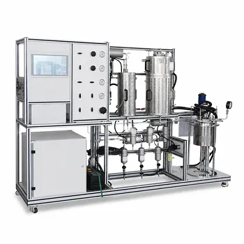
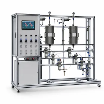
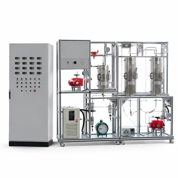
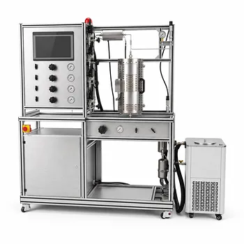
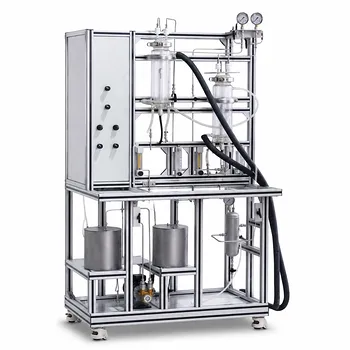
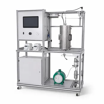
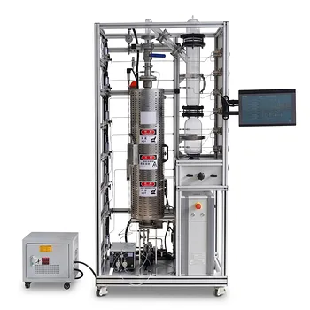
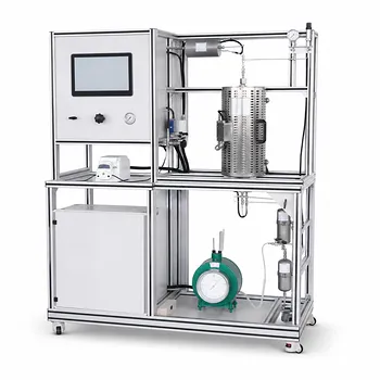
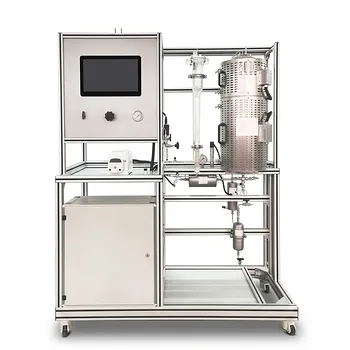
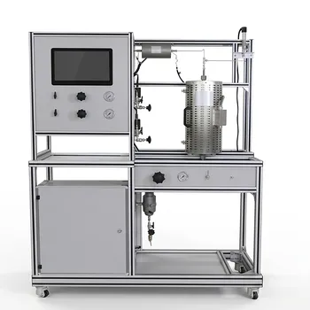
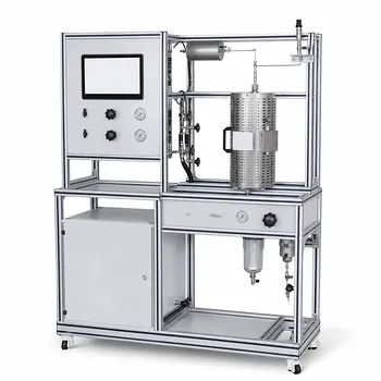
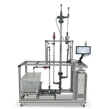
Transitioning from catalyst characterization to scalable reactor operations requires robust, reliable hardware. LABPARK provides premium Educational and Vocational Unit Operations Pilot Plants in chemical engineering, bioprocess & biotech, and environmental & water treatment for universities, research institutes, and enterprises.
Our pilot systems are designed to help you accurately validate kinetic models, control pressure drops, and scale up chemical processes with confidence.
Contact LABPARK today to discover how our pilot plants can elevate your research and training programs!
Related Products
- Multi Functional Catalytic Reaction and Reactor Evaluation Educational Unit Operations Pilot Plant
- Fixed-Bed Chemical Reaction and Gas Dust Tar Removal Unit Operations Pilot Plant
- Crude Benzene Hydrogenation Educational Unit Operations Pilot Plant
- Methanol Synthesis and Catalyst Performance Evaluation Educational Unit Operations Pilot Plant
- Photocatalytic Membrane Separation and Degradation Unit Operations Pilot Plant
People Also Ask
- What reaction engineering principles are shown in a catalytic reactor pilot plant? SO2 Oxidation Guide
- Why is FTIR integration in catalytic pilot plants important? Real-Time Student Insights
- How to analyze active metal distribution & identify catalyst poisoning in pilot plants? Expert Diagnostic Guide
- How do temperature limits and WHSV influence catalytic reactor optimization? Scale-Up Guide
- Why is XPS Critical for Catalytic Reactor Pilot Plants? Optimize Catalyst Performance