Laser Raman spectroscopy offers a powerful, non-destructive fingerprint of catalyst structure, but it can literally cook or disguise the very chemistry you’re trying to see. A robust measurement strategy must therefore neutralize two pervasive foes: thermal decomposition from the laser itself and fluorescence that drowns out the weak Raman signal. You prevent decomposition by continuously moving the sample or the beam to distribute heat density, and you defeat fluorescence by removing the fluorescing contaminants (via oxidation or chemical cleaning) or by exploiting the temporal difference between Raman and fluorescence emission.
Catalyst Raman analysis walks a fine line—enough laser power to generate a signal without destroying the sample. The core solution hinges on thermal load distribution (rotating or scanning the sample) for decomposition and a choice between sample purification (burning off fluorescing organic impurities in O₂ or using chemical oxidizers) and time-gated detection to extract Raman photons before the slower fluorescence overwhelms the detector.
Guarding Against Laser-Induced Decomposition
The high-intensity continuous-wave lasers used in Raman systems (Argon, Krypton, Helium-Neon) dump significant energy into a microscopic spot. For heat-sensitive catalysts, especially those with high surface area or hydrated structures, this local heating quickly surpasses safe thresholds.
Distributing Thermal Energy Across the Sample
The simplest and most universal precaution is to prevent the laser from dwelling on a single point.
Rotating the sample holder is the workhorse technique for pellets and self-supporting wafers. By spinning the sample, the laser effectively illuminates a ring, drastically reducing the time-averaged power density at any one location.
Scanning the laser beam across a static sample surface achieves the same principle. This is particularly useful for irregularly shaped fragments or samples that cannot be spun without risking disintegration. Both methods ensure that heat dissipates before it triggers a phase change or burns the catalyst’s active components.
Matching the Strategy to the Sample Form
Not all catalysts are equally forgiving. Metal-supported catalysts can act as a heat sink, often tolerating higher powers, while pure oxide supports like silicates or aluminas are more prone to local hot-spots. If you observe a visible color change, a darkening spot, or a rapid rise in the background continuum, you are already witnessing thermal damage. Reducing laser power is a complementary measure, but only up to a point—below a certain threshold the Raman signal becomes too weak to detect. Distributing the heat is often the only way to stay above that threshold without destroying the sample.
Banishing Fluorescence from Your Spectra
Fluorescence is the arch-nemesis of Raman spectroscopy. Even trace amounts of organic contaminants—adsorbed on the catalyst surface during synthesis, from storage containers, or from the support material itself—can produce a broad, intense luminescence that completely swamps the sharp Raman lines.
Sample Purification: Destroying the Source
The most direct approach is to eliminate the fluorophores before you even place the sample under the laser.
Heating the sample in an oxygen (O₂) atmosphere to roughly 400–500 °C burns off organic residues, leaving a clean surface. This works beautifully for robust inorganic catalysts that are already calcined at similar temperatures. However, you must ensure the treatment does not alter the phase or sinter the active metal particles—what you gain in signal clarity you can lose in relevance to the actual catalytic state.
Oxidative chemical treatments using hydrogen peroxide (H₂O₂) or concentrated nitric acid offer a lower-temperature alternative. They chemically digest the organics, but residual acids or peroxide can potentially modify surface acid sites or leach mobile species. Always rinse thoroughly and re-dry the sample, and be aware that this aggressive cleaning may be unsuitable if you need to preserve adsorbed intermediates deliberately.
Time-Resolved Detection: A Photonic Shortcut
When you cannot alter the sample without losing critical information—for instance, when studying coked catalysts, adsorbed reaction intermediates, or moisture-sensitive materials—time-gated detection becomes invaluable.
Raman scattering is an instantaneous process (picoseconds), while fluorescence emission has a characteristic lifetime typically in the nanosecond range. By using a pulsed laser and a gated detector (such as an intensified CCD), you can collect only those photons that arrive during the short instant of the laser pulse, effectively cutting off the majority of the longer-lived fluorescence. This technique requires specialized equipment and is not available on all standard Raman microscopes, but it preserves the sample in its native, working state.
Understanding the Trade-offs and Pitfalls
No single solution is a silver bullet. The most common mistake is to chase a "clean" spectrum at the expense of chemical truth.
- Thermal pretreatment can sanitize your data into irrelevance. Burning off coke or reaction intermediates defeats the purpose of in situ or spent-catalyst analysis. Always ask whether the species you are removing is precisely what you wish to measure.
- Rotating or scanning cannot always save a dark-colored sample. Highly absorptive catalysts (e.g., carbon-supported metals) will still heat up rapidly because they convert a large fraction of photons to heat, even if the beam is moving. For these, you may need to combine rotation with drastic power reduction and longer acquisition times.
- Chemical oxidation can introduce new problems. Concentrated nitric acid, for example, can leave behind nitrate residues that themselves fluoresce or decompose under the laser beam.
- Time-gated detection is equipment-intensive and can reduce throughput. The pulsed lasers and gated CCDs are more costly, and if your fluorescence lifetime is extremely short (borderline picosecond scale), the technique offers diminishing returns. It also often requires longer total data collection to achieve comparable signal-to-noise ratios.
Making the Right Choice for Your Goal
Your operational protocol should align directly with what you need to learn from the catalyst. Here’s how to navigate the decision tree:
- If your primary focus is preserving the catalyst in its exact post-reaction state (coke, adsorbates, and all): Employ a sample rotation stage and explore time-resolved detection if available. Minimize laser power to the barely-sufficient level, and expect to use extended acquisition times.
- If your primary focus is the structural integrity of the inorganic matrix (phase, support crystallinity) and organics are just a nuisance: Pre-treat in flowing O₂ at a temperature matching the original calcination step. This often delivers the crispest, fastest spectra.
- If your primary focus is a wide range of oxide or supported metal catalysts on a budget (no pulsed laser option): Start with sample rotation and a thorough chemical clean (H₂O₂ is milder, HNO₃ is more aggressive). Test a small portion to confirm the treatment does not alter the Raman bands you depend on.
- If your primary focus is carbonaceous or deeply colored samples where heating is unavoidable: Abandon the "no damage" ideal and instead bracket your laser power—measure at incrementally lower powers until spectral features stop changing. That defines your safe operating regime, and you accept that some high-power data is compromised.
Your Raman spectrum will only ever be as trustworthy as the precautions you take to capture it without altering the very chemistry it reveals.
Summary Table:
| Challenge | Strategy | How It Works | Best For |
|---|---|---|---|
| Thermal Decomposition | Sample Rotation or Scanning | Distributes laser energy over a larger area to reduce local heat. | Pellets, wafers, and heat-sensitive oxides. |
| Thermal Decomposition | Laser Power Reduction | Lowers total energy input at the cost of signal strength. | Highly absorptive or dark samples. |
| Fluorescence Interference | Thermal/Chemical Oxidation | Burns off or digests organic impurities before analysis. | Robust inorganic catalysts (stable at high temps). |
| Fluorescence Interference | Time-Resolved Detection | Uses pulsed lasers and gated detectors to capture fast Raman photons. | Spent catalysts, coked samples, and active intermediates. |
Elevate Your Chemical Engineering Laboratory with LABPARK
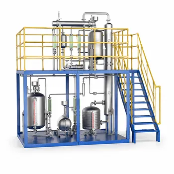
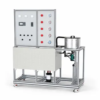
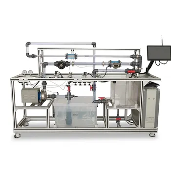
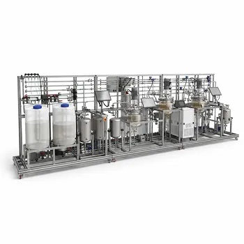
Precise analytical techniques require a solid foundation in experimental design. LABPARK provides advanced Educational and Vocational Unit Operations Pilot Plants in chemical engineering, bioprocess & biotech, and environmental & water treatment designed specifically for universities, research institutes, and enterprises.
Equip your students and researchers with hands-on, industry-standard equipment to master complex chemical processes. Contact us today to discover how LABPARK can customize the perfect pilot plant solution for your lab.
Related Products
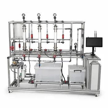
- Multifunctional Membrane Separation Educational Pilot Plant with Ultrafiltration, Nanofiltration, Reverse Osmosis
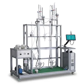
- Continuous Batch Extractive Distillation Educational Pilot Plant
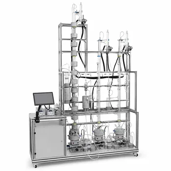
- Multi-Functional Special Distillation Educational Pilot Plant
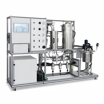
- Multi Functional Catalytic Reaction and Reactor Evaluation Educational Unit Operations Pilot Plant
- Multi-Functional Membrane Separation Educational Pilot Plant for Unit Operations Lab
People Also Ask
- How do PEI, PVDF, and PSU membranes compare in pilot plants? Find the best fit.
- Pervaporation vs. Vapor Permeation: Handling Suspended Solids in Membrane Pilot Plants
- What are the trade-offs of polymer vs inorganic membranes in teaching pilot plants? Key laboratory comparison.
- How can operators optimize operating conditions and cleaning protocols in membrane separation pilot plants to mitigate fouling?
- What are the advantages of membrane separation in pilot plants? Efficient Organic Vapor Recovery