Depth profiling combined with surface analysis turns a catalyst pellet from a "black box" into a three-dimensional map of active metals. By physically removing atomic layers with an argon ion gun and then analyzing the freshly exposed surface, researchers can directly see whether nickel is buried at an interface or concentrated on the outermost shell—data that directly explains mass transfer behavior in pilot reactors. Surface microscopy techniques then complement this by tracking how metal concentrations change across the pellet’s radius, making it possible to link preparation steps to the final performance inside the pore network.
Understanding the spatial fate of active metals is not just analytical curiosity—it’s the key to controlling reaction kinetics, catalyst lifetime, and process economics in unit operations. Depth profiling reveals the vertical layering near pore mouths, while surface analysis maps radial gradients, together giving engineers a complete picture of where metals end up and why that placement matters.
The Critical Link Between Metal Location and Catalyst Performance
Why Spatial Distribution Determines Reactor Behavior
A catalyst’s effectiveness in a unit operation reactor depends on whether reactant molecules can reach the active sites. If most of the nickel or molybdenum sits deep inside narrow pores, diffusion limitation can starve the reaction, while an excess on the external surface might cause rapid coking or metal leaching.
Engineers need to know exactly where the metals are, not just how much is present. That’s because the impregnation sequence, drying conditions, and calcination all influence whether the active phase forms a thin crust, a uniform egg-shell, or an egg-yolk profile. Each pattern brings a different balance of activity, selectivity, and stability.
The Cost of Ignoring Spatial Distribution
When pilot-scale hydrotreating catalysts deactivate prematurely, the root cause often traces back to an uneven metal distribution. For example, contaminants like vanadium can accumulate in certain pore regions, as revealed by elemental mapping. Without spatial insight, operators waste time adjusting temperature and pressure instead of fixing the catalyst loading method.
How Depth Profiling Unveils Subsurface Distributions
Ar-Ion Sputtering as a Subsurface Probe
Depth profiling works by bombarding the sample with an argon ion beam that slowly erodes away surface atoms. After each etching step, a surface-sensitive technique like XPS (X-ray photoelectron spectroscopy) measures the remaining composition.
This process creates a composition-versus-depth profile, showing whether nickel resides just below the surface, at the molybdenum-alumina interface, or deeper within the support. For NiMo/Al₂O₃ precursors, such depth profiles have revealed that the order of impregnation dramatically changes the nickel location—a fact that pilot-scale preparation protocols can then exploit to tune catalyst activity.
Connecting Depth Data to Pore Structure
The data from sputtering are not just chemical snapshots; they reflect the interaction between precursor solution and pore geometry. A sharp concentration drop after a few nanometers suggests metal retention near pore entrances, while a slow decline points to deeper penetration. This information lets unit operations researchers predict whether a given catalyst will suffer from pore mouth plugging or efficient internal surface utilization.
Surface Analysis: Mapping the Pellet’s Radial Landscape
Microscopy Techniques for Concentration Profiles
While depth profiling investigates the vertical axis near a pellet’s exterior, surface analysis microscopy—such as EPMA, SEM, and STEM—provides the lateral and radial picture. These tools scan across a catalyst cross-section to measure how the active metal concentration changes from the edge to the center.
For hydrotreating catalysts, such maps can clearly show a vanadium contamination ring that builds up at a specific radial depth, directly linking pore size to metal deposition and deactivation. This insight is vital for choosing carriers with the right pore architecture for a given feedstock.
The Power of Relative Intensity Ratios
Absolute quantification on rough, porous surfaces is unreliable. Instead, researchers rely on the corrected integrated intensity ratio of metal to support signal (Imetal/Isupport). If this ratio is higher than predicted by the nominal metal surface density, it indicates enrichment on the external surface—a sign of metal migration during drying. A ratio lower than expected points to sintering or to metals hidden inside small pores that are not accessible to the analysis beam. This simple metric quickly flags whether a preparation method is pushing metals to the wrong locations.
Combining the Tools for a Complete 3D Picture
Integrating Vertical and Radial Information
No single technique can fully capture the complex distribution in a porous pellet. Depth profiling reveals the layered composition near the outer surface, while EPMA or STEM provides radial concentration gradients. Together, they answer the central question: Are the active metals positioned where the reaction can actually use them?
For instance, depth profiling might show a nickel-rich skin, and radial mapping might confirm that the same skin extends uniformly around the pellet. That combined dataset immediately tells the engineer that the catalyst will be highly sensitive to external mass transfer, guiding the choice of reactor space velocity.
Using the Dual Approach for Catalyst Development
When developing a new impregnation protocol, researchers can iterate rapidly: prepare a sample, use depth profiling to check interface sharpness, and then use line scans from SEM-EDS to verify radial uniformity. This feedback loop shortens development time and reduces the number of expensive pilot-scale tests, because the nanoscale metal layout is no longer a mystery.
Understanding the Trade-offs and Limitations
Depth Profiling Challenges in Porous Systems
The complex pore network and surface roughness introduce uncertainty in depth resolution. The ion beam etches at different rates on sharp edges and within crevices, causing the apparent depth to mix signals from different real depths. The result is a smeared profile that requires careful interpretation—trends are reliable, but absolute depth values are not.
Additionally, preferential sputtering can alter the surface composition during analysis, so the measured ratios may not perfectly represent the true distribution. Researchers must use standard corrections and compare relative changes rather than absolute numbers.
Surface Analysis Constraints
Microscopy methods like EPMA offer spatial resolution down to a micrometer, but the electron beam excites a relatively large volume in porous materials, again blurring the exact location. Moreover, analyzing the pellet interior requires mechanical sectioning, which can smear metals across the surface and create artifacts. The relative intensity ratio method avoids many of these pitfalls but only provides an indirect inference of pore-level distribution rather than a direct image.
How to Apply These Insights to Your Research
Select the approach based on the specific engineering question you need to answer.
- If your primary focus is optimizing the impregnation sequence: Use depth profiling with Ar-ion sputtering to identify the exact interface where active metals concentrate. Adjust the order and drying rate to shift that interface for better activity.
- If your primary focus is preventing rapid deactivation in a pilot reactor: Combine cross-sectional EPMA or SEM-EDS with relative intensity ratio analysis. Monitor for surface enrichment or a ring of contaminants that points to pore plugging or metal migration.
- If your primary focus is scaling up a new catalyst formulation: Validate that the metal distribution remains consistent from lab-scale samples to production batches by correlating depth profiles and radial maps. A change in distribution will alert you to scale-up issues long before a full reactor run fails.
- If your primary focus is selecting a support with the right pore size: Map contaminant profiles across the pellet radius using STEM and compare the depth of penetration. Choose the pore diameter that keeps deposition within a safe zone, extending catalyst life.
By turning analytical data into a clear spatial map of active metals, you move from trial-and-error preparation to deliberate design—giving you control over every cubic nanometer of the catalyst’s interior.
Summary Table:
| Technique | What It Measures | Value for Unit Operations | Key Limitations |
|---|---|---|---|
| Depth Profiling (XPS + Ar Sputtering) | Vertical composition layering near pore mouths | Optimizes impregnation sequence & checks interface sharpness | Roughness causes profile smearing |
| Surface Analysis (EPMA / SEM / STEM) | Radial concentration gradients across pellet cross-section | Detects contaminant accumulation & migration during drying | Mechanical sectioning can create surface artifacts |
Scale Up Your Chemical Engineering Research with LABPARK
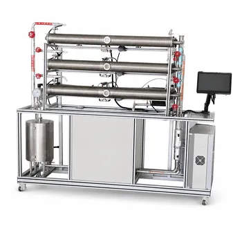
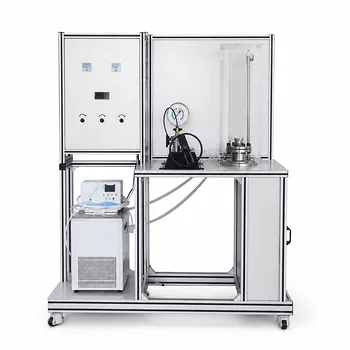
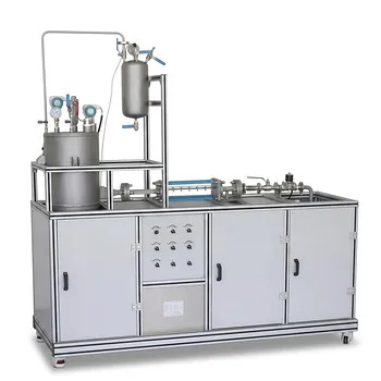
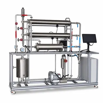
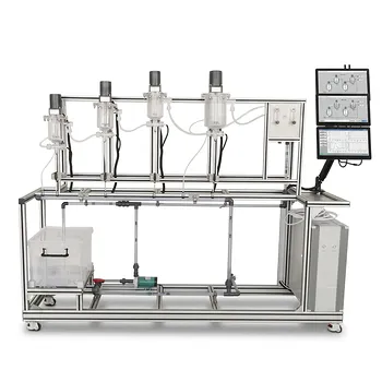
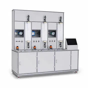
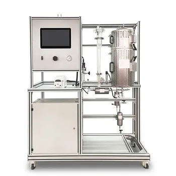
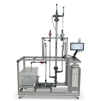
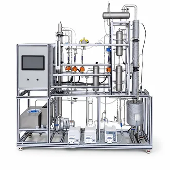
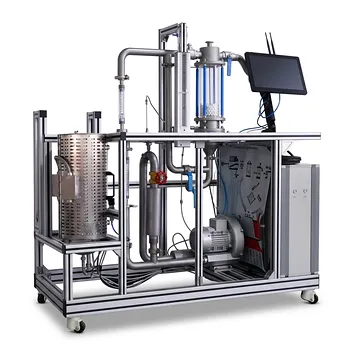
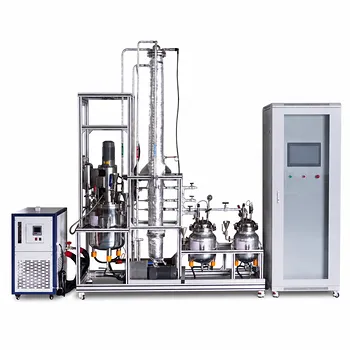
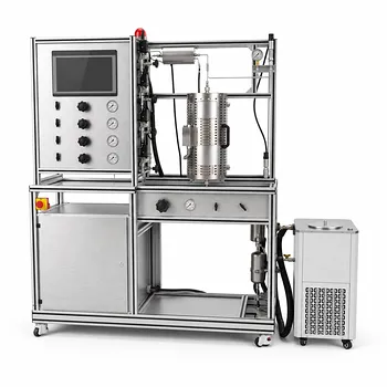
Translating microscopic catalyst insights into macroscopic reactor success requires robust pilot-scale validation. LABPARK provides state-of-the-art Educational and Vocational Unit Operations Pilot Plants in chemical engineering, bioprocess & biotech, and environmental & water treatment.
Designed specifically for universities, research institutes, and enterprises, our pilot plants allow you to simulate real-world kinetics, mass transfer, and deactivation profiles under controlled conditions.
Ready to elevate your research capabilities? Contact LABPARK today to find the perfect pilot plant solution for your lab!
Related Products
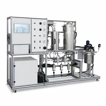
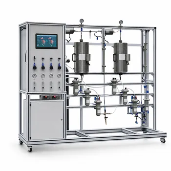
- Multi Functional Catalytic Reaction and Reactor Evaluation Educational Unit Operations Pilot Plant
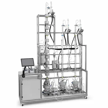
- Continuous Batch Extractive Distillation Educational Pilot Plant
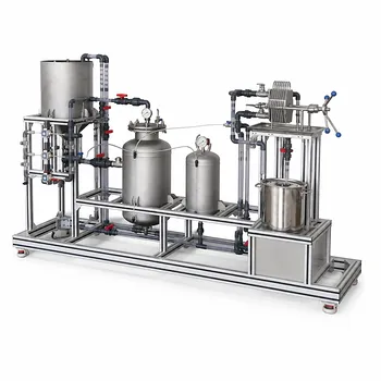
- Constant Pressure Filtration Educational Unit Operations Pilot Plant
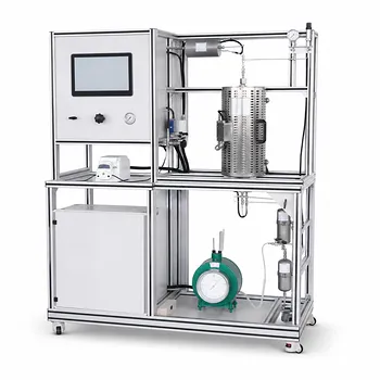
- Educational Unit Operations Pilot Plant for Intraparticle Diffusion Effective Factor Measurement
- Three-Tube Heat Transfer Educational Pilot Plant for Unit Operations Training
People Also Ask
- What reaction engineering principles are shown in a catalytic reactor pilot plant? SO2 Oxidation Guide
- Why is FTIR integration in catalytic pilot plants important? Real-Time Student Insights
- How to analyze active metal distribution & identify catalyst poisoning in pilot plants? Expert Diagnostic Guide
- How do temperature limits and WHSV influence catalytic reactor optimization? Scale-Up Guide
- What operational insights do catalyst pellet concentration profiles provide? Optimize Reactor Yield