Start by understanding that the question isn't IR or Raman—it's about how to use them as complementary probes for different parts of the catalytic puzzle.
For a catalytic reaction engineering pilot plant, the decision hinges on two things: the chemical nature of your catalyst and the specific bonds you need to observe. Raman spectroscopy excels at seeing the catalyst backbone itself and symmetric, non‑polar vibrations of adsorbed species, all while being virtually invisible to the strong scatter from common oxide supports like alumina and silica. Infrared (IR) spectroscopy, by contrast, is the go‑to technique for polar functional groups, acidic sites, and studying the dynamics of surface intermediates under transient reaction conditions.
Core Takeaway
The choice between IR and Raman in catalyst characterization is not about which method is “better” but about which physical information each technique can provide. Raman reveals the metal–oxygen framework and symmetric adsorbate modes without support interference; IR identifies polar reaction intermediates, probes acidic sites, and readily supports transient kinetics. In practice, a comprehensive understanding of catalytic mechanism demands both—deployed strategically based on the problem you're solving.
Why the Two Techniques See Different Chemistry
The Mutual Exclusion Principle Sets the Ground Rules
IR and Raman probe the same molecular vibrations but through completely different physical mechanisms.
IR spectroscopy measures the absorption of light and requires a change in the molecule’s dipole moment during vibration.
Raman spectroscopy relies on inelastic scattering of light and requires a change in polarizability—the ability of the molecule’s electron cloud to be distorted.
This leads to a critical selection rule: for molecules with a center of symmetry, mutual exclusion applies. Any vibrational mode active in IR is forbidden in Raman, and vice versa.
Even for non‑centrosymmetric molecules, the two techniques often highlight different parts of the spectrum.
In practical terms, symmetric stretches and non‑polar bonds (like C=C, S–S, Mo=O) give strong Raman signals, while asymmetric stretches and polar groups (C=O, O–H, N–H) dominate IR spectra.
Knowing this lets you decide upfront which tool will see the species you care about.
Key Decision Factors for Catalyst Characterization
The Nature of the Catalyst Support
A decisive advantage of Raman in pilot plant settings is its insensitivity to common oxide supports.
Alumina (Al₂O₃) and silica (SiO₂)—the backbone of many industrial catalysts—are notoriously strong IR absorbers that can obscure the low‑frequency region where metal–adsorbate bonds appear.
In Raman spectroscopy, these supports produce only weak, broad features, leaving the catalyst’s own vibrational modes and metal–adsorbate bands clearly visible.
This means that if your goal is to study the catalyst structure itself—for example, the Mo–S linkages in a hydrodesulfurization (HDS) catalyst or the active oxygen species on a vanadia catalyst—Raman is the unambiguous first choice.
You can directly observe the metal–oxygen framework below 1000 cm⁻¹ without drowning in support background.
The Bond You Wish to Observe: Polar vs. Non‑Polar
IR spectroscopy’s strength is the detection of polar functional groups and the ability to follow how they change during reaction.
When you need to monitor carbonyl (C=O) stretches, hydroxyl (O–H) groups, or adsorbed CO, IR delivers sharp, concentration‑sensitive peaks.
For example, differentiating Brønsted acid sites from Lewis acid sites on a solid acid catalyst is traditionally done by IR analysis of adsorbed pyridine or ammonia—a task Raman cannot easily replicate with the same clarity.
Conversely, symmetric vibrations and non‑polar bonds often vanish in IR but shine in Raman.
Studying the symmetric breathing mode of an adsorbed aromatic molecule or the Mo=O terminal oxo bond in an HDS catalyst is best done with Raman.
If you’re primarily interested in the catalyst’s own lattice vibrations or symmetric adsorbate structures, lean towards Raman.
Accessing Low‑Frequency Modes and Metal–Adsorbate Bonds
The low‑frequency region (below ~600 cm⁻¹) contains the metal–adsorbate stretching and bending modes that are direct fingerprints of the catalytic act.
Standard mid‑IR optics and the strong absorption of supports make this region difficult to access with IR.
Raman, especially with a single‑monochromator system, easily reaches down to 100 cm⁻¹, revealing vibrations like Pt–C, Pd–O, or Mo–S.
For pilot plant work where you want to watch the actual bond between catalyst and reactant in real time, Raman’s low‑frequency access is a compelling advantage.
Practical Considerations in a Pilot Plant Environment
Fluorescence Can Kill a Raman Signal
Raman’s biggest weakness is fluorescence interference.
Many organic molecules, catalyst residues, or even the support itself can fluoresce strongly, swamping the much weaker Raman scattering.
If your reaction stream contains heavy hydrocarbons, colored species, or trace impurities, a Raman spectrum may be completely buried under a fluorescence background.
The remedy lies in laser wavelength selection. Shorter wavelengths (532 nm) give high Raman intensity but trigger more fluorescence; longer wavelengths (785 nm, 1064 nm) dramatically reduce fluorescence but generate lower signal and risk sample heating.
In a pilot plant, if you anticipate fluorescent species—for example, during biomass conversion or coking studies—plan to use a 785 nm or 1064 nm Raman system and be prepared for longer acquisition times or the use of highly sensitive InGaAs detectors.
Water and CO₂ Interfere Strongly in IR
IR spectroscopy is exceptionally sensitive to water vapour and dissolved carbon dioxide, which produce strong, sharp absorption bands around 3400 cm⁻¹ (O–H stretch), 1640 cm⁻¹ (H–O–H bend), and 2350 cm⁻¹ (CO₂).
In a flow reactor or in‑situ cell, any trace of moisture will pollute the spectrum.
Operationally, you must purge the spectrometer with dry air or nitrogen and ensure sample streams are anhydrous, or use subtraction algorithms—a non‑trivial overhead in a busy pilot plant.
In contrast, water is a very weak Raman scatterer, making Raman the superior choice for aqueous‑phase reactions or when studying catalysts under humid conditions.
If your catalytic process involves liquid water or you cannot avoid moisture, Raman’s tolerance is a major practical advantage.
Path Length and Sampling Depth: Mid‑IR vs. NIR
Conventional mid‑IR spectroscopy of liquids demands extremely short path lengths (≤0.1 mm) because fundamental vibrations absorb so strongly.
In a flowing pilot plant stream, this means you’ll likely need an ATR (Attenuated Total Reflectance) probe to handle particulates, bubbles, and to control the sampling depth.
Near‑IR (NIR) spectroscopy, which measures weaker overtones, allows much longer path lengths (1–100 mm) and is therefore easier to interface with process lines—but it requires multivariate chemometrics for interpretation and is not suitable for gas‑phase or low‑concentration monitoring.
For gas‑phase heterogeneous catalysis in a pilot reactor, mid‑IR (or FTIR) is the standard; for liquid‑phase monitoring, Raman with a fiber‑optic probe often provides a simpler, more robust solution.
Transient Kinetics and Isotopic Labelling: IR’s Time‑Domain Edge
If your research goal is to identify reaction intermediates and measure their surface lifetimes, IR spectroscopy offers a well‑established platform for transient experiments.
By using fast‑scanning FTIR spectrometers (0.1–1 second time resolution) and perturbing the reactor with pulses, steps, or isotopic switches (e.g., D₂ or ¹³C), you can directly correlate the rate of band disappearance with the overall reaction rate.
This capability is critical when scaling up a new catalytic process, because it tells you which adsorbed species are true kinetic intermediates and which are mere spectators.
Raman can also be used time‑resolved, but its inherently weaker signal and potential fluorescence background can make rapid transient studies more challenging unless you have an optimized, high‑sensitivity system.
Understanding the Trade‑offs and Common Pitfalls
The “Only‑One‑Technique” Trap
A frequent mistake is attempting to solve every characterization problem with a single technique.
Researchers who rely solely on IR may never see the symmetry‑forbidden vibrations that hold the key to the active site structure, and they will struggle to probe the low‑frequency region.
Those who choose only Raman may miss the polar intermediates (such as surface carboxylates or alkoxides) that IR reveals so clearly.
The two techniques are complementary, not competitive.
Pilot plants that invest in both—or at least design flexible optical access for both—gain a far more complete picture of catalyst state and surface chemistry.
The Fluorescence‑vs‑Signal Intensity Trade‑off
In Raman, there is no free lunch. Shorter wavelength lasers (532 nm) give strong signals and fast acquisitions, ideal for real‑time monitoring of rapid processes.
But if your catalyst or reaction mixture fluoresces even slightly, that signal is lost.
Longer wavelength lasers (785 nm, 1064 nm) quench fluorescence, but the signal drops dramatically (intensity ∝ 1/λ⁴), and you may need a higher‑power laser that risks thermally altering the sample.
Always test your specific catalyst‑reaction system under realistic conditions before finalizing the Raman configuration.
Confusing NIR with Mid‑IR Functionality
Near‑IR and mid‑IR differ fundamentally in the type of spectral information they provide.
Mid‑IR sees fundamental vibrations and gives clear molecular fingerprints, making it ideal for catalytic surface studies.
NIR sees overtones and combinations; while NIR is a powerful process analytical tool for bulk liquid composition, it cannot directly distinguish surface‑adsorbed species on a catalyst in the way mid‑IR can.
For characterizing adsorbed intermediates, stick with mid‑IR (or Raman). Reserve NIR for bulk compositional monitoring of the liquid feed or product stream.
Ignoring Sample Preparation and Optical Access
Both techniques demand careful optical design.
IR requires sample presentation that avoids peak saturation (target ~90% baseline transmittance) and eliminates free water and CO₂.
Raman needs the laser to focus cleanly on the catalyst bed or flowing sample without bubbles or moving particles; a poorly aligned probe can collect wall signal rather than the reaction zone.
In a pilot plant, engineering the right in‑situ cell or fiber‑optic probe is often as important as choosing the spectrometer itself.
Making the Right Choice for Your Pilot Plant
Your decision should flow from the exact scientific question you’re asking and the practical constraints of your reactor environment. Use the following goal‑oriented guidelines to select the appropriate approach.
- If your primary focus is the catalyst structure and low‑frequency metal–adsorbate bonds: Start with Raman. Its freedom from support interference and access down to 100 cm⁻¹ directly reveal the active phase and how reactants bind to it.
- If your primary focus is distinguishing adsorbed polar intermediates or acid‑site properties: Choose mid‑IR. The sensitivity of IR to dipole‑moment changes makes it the method of choice for tracking carbonyls, carboxylates, and Brønsted/Lewis acid sites via probe molecules like ammonia or pyridine.
- If your catalytic process involves aqueous media or moisture‑sensitive steps: Favor Raman. Water is a very weak Raman scatterer and will not drown out your catalyst–adsorbate signals, whereas it severely interferes in IR.
- If your goal is to measure surface reaction kinetics and identify short‑lived intermediates: Use mid‑IR with a fast FTIR system. Combine it with transient feeds and isotopic labelling to separate true intermediates from spectators and to derive kinetic parameters for scale‑up.
- If your feedstocks are inherently fluorescent (e.g., heavy oils, biomass, polymers): Opt for Raman with a 785 nm or 1064 nm laser, or stick with IR if the fluorescence is too severe. Always validate that the chosen Raman wavelength gives clean spectra under actual reaction conditions.
- If you are monitoring bulk liquid composition and catalyst characterization is secondary: A combination of NIR (for bulk stream analysis) and Raman or mid‑IR (for surface‐species studies) will give the most complete picture.
Ultimately, the most successful pilot plant strategy is to build optical access for both Raman and IR from the start. The investment pays off by providing independent, cross‑validating views of the catalyst at work—a capability that is no longer a luxury when you are tasked with scaling up a catalytic process with confidence.
Summary Table:
| Feature / Need | Infrared (IR) Spectroscopy | Raman Spectroscopy |
|---|---|---|
| Physical Mechanism | Light absorption (requires dipole change) | Inelastic scattering (requires polarizability change) |
| Best For | Polar groups (C=O, O-H), acid sites, transient kinetics | Non-polar/symmetric bonds (C=C, Mo=O), low-frequency modes |
| Support Interference | Strong absorption by oxide supports (SiO₂, Al₂O₃) | Minimal interference; catalyst backbone remains visible |
| Water Sensitivity | High interference (requires dry environments) | Very low interference (ideal for aqueous-phase reactions) |
| Main Drawback | Difficult to access low-frequency (<600 cm⁻¹) region | High risk of fluorescence interference from organic residues |
Optimize Your Chemical Engineering & Catalysis Research
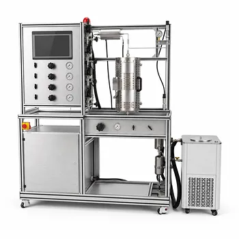
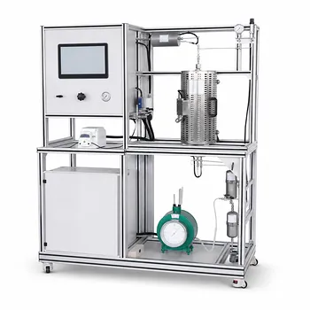
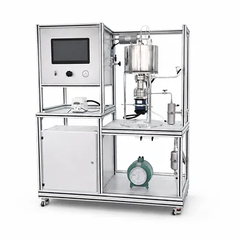
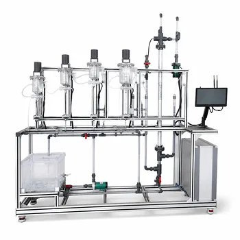
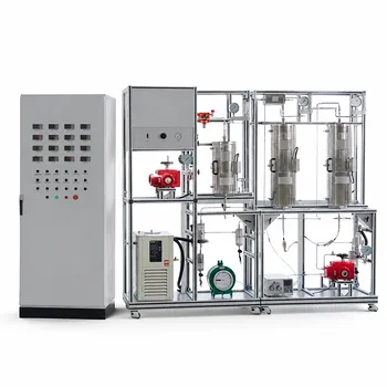
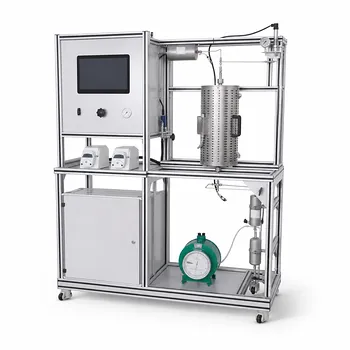
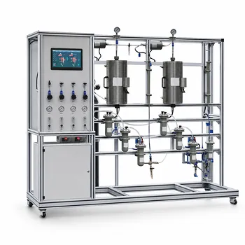
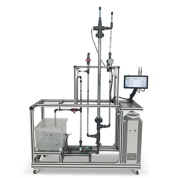
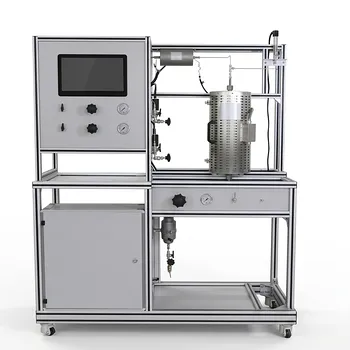
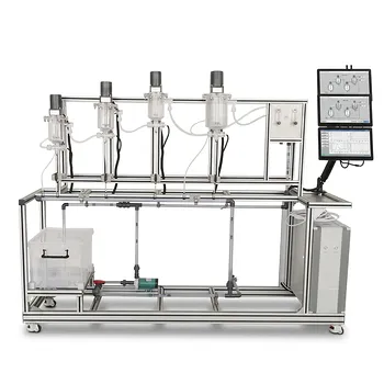
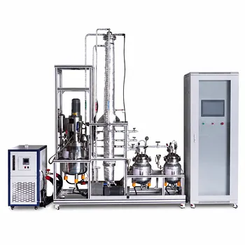
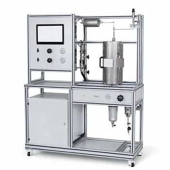
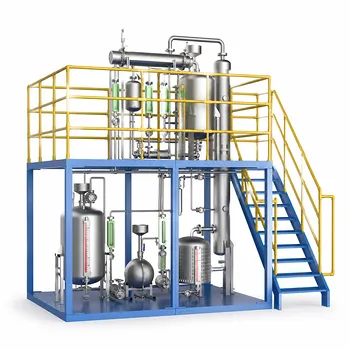
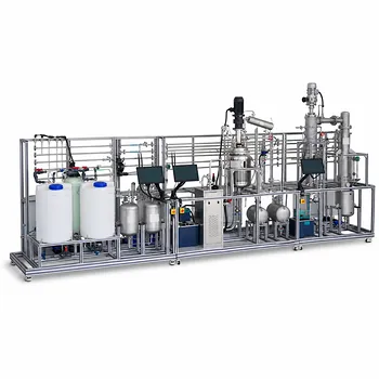
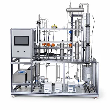
Setting up a catalysis laboratory or scaling up to a pilot plant requires the right integration of analytical techniques and reactor systems. LABPARK provides advanced Educational and Vocational Unit Operations Pilot Plants in chemical engineering, bioprocess & biotech, and environmental & water treatment. We help universities, research institutes, and enterprises design and equip custom pilot setups tailored to precise research goals.
Ready to build or upgrade your catalytic reaction engineering pilot plant? Contact the LABPARK experts today to discuss your system requirements and receive a tailored solution.
Related Products
- Methanol Synthesis and Catalyst Performance Evaluation Educational Unit Operations Pilot Plant
- Educational Unit Operations Pilot Plant for Intraparticle Diffusion Effective Factor Measurement
- Micro-Scale Gas-Solid Catalytic Reaction Educational Pilot Plant
- Internal Circulation Gradient Free Catalytic Reaction Educational Pilot Plant
- Fluidized Bed Gas Solid Catalytic Reaction Educational Pilot Plant
People Also Ask
- Why is a purge system necessary when operating a gas recirculation loop in a methanol synthesis pilot plant? (Guide)
- How do temp & pressure affect methanol synthesis pilot plants? Optimize equilibrium and catalyst performance.
- Why do modern methanol pilot plants operate at lower pressures? Catalyst & Feed Requirements Explained
- What are the operational requirements for catalyst activation? Safe Methanol Pilot Plant Operation
- Why Compare Predicted and Experimental Excess Enthalpy? Key to Accurate Pilot Plant Scale-up