Non-specific binding is one of the most persistent challenges in fiber-optic biosensing. In biotechnology pilot systems, it creates a fog of background noise that can mask the true signal from your target analyte, leading to inaccurate process monitoring and control decisions. The core methods to tackle this are a three-pronged approach: surface passivation with blocking agents, intelligent selection of fluorescent tracers, and calibration-based subtraction using control fibers. Each method either physically prevents unwanted adhesion or mathematically corrects for what remains, ensuring that the final measurement reflects only the specific interaction you care about.
Non-specific binding is minimized primarily by coating the optical fiber surface with blocking agents (like casein or non-target IgG) and by optimizing the probe design to select tracers with inherently low non-specific affinity. Any residual signal is then corrected by subtracting the response of a control fiber during calibration. This integrated strategy turns a noisy sensor into a reliable quantitative tool.
The Hidden Cost of Non-Specific Binding in Pilot Systems
Pilot-scale bioprocessing demands real-time, accurate concentration data. When a fluorescent tracer or the analyte itself sticks to places it shouldn’t—the quartz fiber surface, stray proteins, or even the blocking layer—the sensor reports a composite signal. That composite distorts true kinetics, wastes valuable material, and can trigger flawed process adjustments.
Why the Fiber Surface is a Magnet for Interference
The silica-based core of an optical fiber carries a slight negative charge and numerous silanol groups. Proteins and many small-molecule tracers readily adsorb via hydrophobic interactions, electrostatic forces, or hydrogen bonding. Even after extensive washing, a stubborn monolayer can remain, generating a persistent background fluorescence that rivals the specific signal, especially at low target concentrations.
The Problem with Protein-Stabilized Formulations
In pilot systems, fermentation or cell culture media often contain stabilizing proteins like bovine serum albumin (BSA) or casein. While these additives protect product quality, they become additional surfaces for fluorescent tracers to bind non-specifically. A tracer designed for a perfect buffer may behave completely differently when a complex protein soup is present, inflating the apparent concentration of your target.
Strategies to Minimize Non-Specific Binding
The first line of defense is to physically block the binding sites before the measurement begins. This is a preventative step that removes the majority of the interference.
Surface Passivation: Coating the Battlefield
The most direct method is to flood the fiber surface with an inert, protein-rich blocking agent. Common choices are casein (a milk protein) or a non-target immunoglobulin G (IgG)—an antibody that has no affinity for the analyte. These molecules adsorb tenaciously to the silica, filling all the high-energy sites and creating a “forest” of harmless proteins. When the fluorescent tracer is introduced, it has far fewer locations to land uninvited, dramatically reducing physical adsorption.
Tracer Optimization: Selecting the Right Spy
Not all fluorescent tracers are created equal; their non-specific “stickiness” is a key selection criterion. During assay development, structurally related tracer candidates (e.g., different fluorophore-antibody conjugates) are tested at low, process-relevant concentrations—often around 10 nM. The goal isn’t just the highest binding affinity for the target, but the highest signal-to-noise ratio. A tracer with slightly lower affinity but dramatically lower non-specific binding will provide cleaner, more reliable data in a pilot system, where optical background drifts over days.
Correcting Residual Non-Specific Signal
Even with rigorous blocking and optimized probes, some non-specific binding can persist, particularly in long-duration runs where surface fouling evolves. The strategy here is to measure the unwanted signal independently and subtract it.
The Control Fiber: A Mirror Without the Target
A dedicated control fiber is prepared identically to the sensing fiber, except it lacks the specific recognition element for the analyte. For example, it might be coated only with the blocking agent or with a mismatched antibody. During the run, both the sensing and control fibers are exposed to the same sample matrix, temperature, and fluorescent tracer. The signal from the control fiber represents the sum of all non-specific interactions. By subtracting this control signal from the sensing fiber’s output, you mathematically isolate the specific binding response, effectively zeroing out the background.
Calibration Integration: Turning Subtraction into Accuracy
The subtraction is not a simple post-processing fix; it’s baked into the calibration curve. Calibration standards are measured with both the sensing and control fibers, establishing a relationship between the net specific signal and the analyte concentration. In operation, the system continuously logs both raw signals and outputs the corrected value. This dynamic correction accounts for gradual drift in non-specific binding that would otherwise shift your baseline over a multi-day pilot fermentation.
Understanding the Trade-offs
These methods are powerful, but they are not without compromises in a real pilot environment.
Blocking Agents Can Become New Targets
Excessive blocking can backfire. A thick, loosely packed layer of casein or IgG may present new binding motifs that tracers actually recognize. The blocking step must be optimized for concentration and incubation time to form a monolayer that saturates the surface without creating a three-dimensional hydrogel that traps the tracer. Additionally, casein’s lot-to-lot variability can impact reproducibility, so quality-controlled preparations are essential.
Tracer Optimization Limits Your Flexibility
Screening multiple tracer candidates for the lowest non-specific binding is time-consuming. A tracer chosen for its superb signal-to-noise at 10 nM might underperform if the target concentration spikes unexpectedly. There’s an inherent trade-off between the ideal “clean” probe in a controlled pilot environment and the robustness needed for a process that may see wide concentration swings.
Subtraction Assumes Linear Additivity
The mathematical correction works best when non-specific binding is independent and additive. If the presence of the target analyte actually suppresses or enhances the amount of non-specific binding (a phenomenon known as matrix effect), a simple subtraction will mis-correct. In those cases, more advanced calibration models or internal standard spiking may be required, adding complexity.
Making the Right Choice for Your Pilot System
Selecting the right combination of methods depends on the constraints of your process and the level of accuracy required.
- If your primary focus is rapid development and low cost: Start with a robust surface blocking protocol using a commercially available casein or protein-free synthetic blocker. Pair it with a tracer that has been prescreened for low background, and rely on a single control fiber for the most critical measurement points.
- If your primary focus is maximum sensitivity at low concentrations (pM–nM range): Invest heavily in tracer optimization. Screen structurally related conjugates specifically in your process media at your target concentration, and use a dedicated control fiber for each sensor. Validate that the subtraction model holds across your entire expected concentration range.
- If your primary focus is long-term stability over multi-day pilot runs: Regularly refresh or regenerate the blocking layer if the sensor design allows, and implement automated, frequent control-fiber readings to correct for progressive fouling. Consider using a non-fouling co-blocking agent like a zwitterionic polymer layer underneath the protein blocker.
By treating non-specific binding not as a single problem but as a layered challenge of prevention and correction, you turn your fiber-optic biosensor into a trustworthy partner for process scale-up.
Summary Table:
| Method | Action / Principle | Key Benefit |
|---|---|---|
| Surface Passivation | Coating fiber with blocking agents (casein or non-target IgG) | Physically blocks unwanted adhesion sites |
| Tracer Optimization | Selecting tracers with low inherent non-specific affinity | Maximizes signal-to-noise ratio in complex media |
| Control Fiber Subtraction | Subtracting control fiber response during calibration | Mathematically corrects for residual background |
Optimize Your Bioprocess Scale-Up with LABPARK
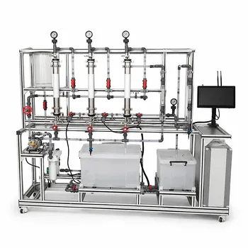
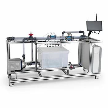
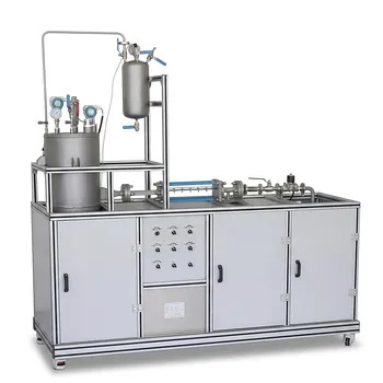
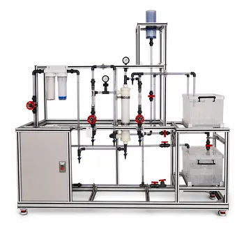
Achieving precise, noise-free monitoring is critical for successful pilot-scale operations. LABPARK provides advanced Educational and Vocational Unit Operations Pilot Plants in chemical engineering, bioprocess & biotech, and environmental & water treatment, specifically tailored for universities, research institutes, and enterprises.
Ready to enhance your lab's capabilities and training efficiency? Contact us today to explore our high-performance pilot plant solutions!
Related Products
- Multifunctional Membrane Separation Educational Pilot Plant with Ultrafiltration, Nanofiltration, Reverse Osmosis
- Orifice and Venturi Flowmeter Calibration Educational Pilot Plant for Fluid Mechanics Laboratory
- Cavitation Phenomenon Demonstration and Analysis Educational Unit Operations Pilot Plant
- Ultrafiltration Membrane Separation Educational Pilot Plant
- Constant Pressure Filtration Educational Unit Operations Pilot Plant
People Also Ask
- Pervaporation vs. Vapor Permeation: Handling Suspended Solids in Membrane Pilot Plants
- How do PEI, PVDF, and PSU membranes compare in pilot plants? Find the best fit.
- What are the trade-offs of polymer vs inorganic membranes in teaching pilot plants? Key laboratory comparison.
- How can operators optimize operating conditions and cleaning protocols in membrane separation pilot plants to mitigate fouling?
- What are the advantages of membrane separation in pilot plants? Efficient Organic Vapor Recovery