Accurate fluorescence polarization (FP) measurements in chemical engineering and bioprocess pilot plants are impossible without determining and applying the G-factor. This instrument-specific correction factor compensates for the inherent bias in your optical system’s transmission of vertically versus horizontally polarized light. Without it, your calculated polarization and anisotropy values are skewed, leading to misinterpretation of molecular binding events and unreliable process control feedback.
The G-factor is not a one-time checkbox but a fundamental calibration against optical bias. In a pilot plant where FP sensors monitor binding kinetics, ignoring it injects systematic error into every reading; calibrating it with a scattering standard under horizontal excitation corrects for unequal monochromator transmission, ensuring your data reflects real molecular behavior, not instrument artifacts.
The Critical Role of the G-Factor in FP Measurements
Correcting Inherent Optical Bias
Every spectrofluorometer has monochromators that exhibit different efficiencies for vertically and horizontally polarized light. This difference arises from the grating’s polarization-dependent reflectivity and other optical components. When you measure the parallel and perpendicular fluorescence intensities of your target analyte, the raw detector signals are distorted by this built-in bias. The G-factor (the system’s polarization response) quantifies this asymmetry and allows you to mathematically correct for it, restoring the true intensity ratio.
Impact on Polarization and Anisotropy Calculations
Fluorescence polarization (P) and anisotropy (r) are ratios of intensity differences to total intensity. If your perpendicular intensity is systematically under- or over-reported due to unequal transmission, both P and r will be erroneous. For example, a G-factor of 1.2 means the system transmits 20% more of one polarization component than the other. Without correction, a stable sample can appear to change its binding state simply because the instrument’s bias varies with wavelength or alignment. Applying the G-factor transforms raw intensities into unbiased measurements, ensuring the molecular information you extract—binding constants, kinetic rates—is accurate.
Why This Matters in Pilot Plant Environments
In a bioprocess or chemical engineering pilot plant, FP sensors are often used to monitor biomolecular interactions such as DNA-protein binding, ligand intercalation, or protein conformational changes. These measurements directly influence process decisions: adjusting feed rates, triggering separations, or validating product quality. A systematic bias left uncorrected can mimic a process shift or mask a real one. Moreover, in a training or research setting, teaching the correct determination of the G-factor instills the discipline of instrument validation—a cornerstone of reproducible analytical unit operations.
How to Calibrate the G-Factor in a Pilot Plant Sensor
The Principle: Using a Scattering Standard
A perfectly depolarizing scatterer isotropically redirects light, so under horizontally polarized excitation its scattered light at 90° consists of equal vertical and horizontal components. Any difference in the detected signals when the emission polarizer is switched must therefore be caused solely by the instrument’s transmission imbalance. A dilute suspension of glycogen (or another non-fluorescent scattering agent) serves as this ideal standard. By measuring the scattered light with the excitation polarizer fixed at horizontal, you can directly calculate the G-factor.
Step-by-Step Procedure
-
Prepare a scattering sample. Use a fresh, dilute glycogen suspension in water buffer that is optically clear but turbid. Ensure it does not fluoresce at your excitation wavelength.
-
Set the excitation polarizer to horizontal. This guarantees the scattered light incident on the emission monochromator is depolarized.
-
Measure the two emission intensities. With the emission polarizer set to vertical, record I_HV. Then rotate the emission polarizer to horizontal and record I_HH. The excitation wavelength should match your target analyte’s excitation, and the emission wavelength should be identical (often the same wavelength as excitation for a pure scatterer).
-
Calculate the G-factor. The formula is:
G = I_HV / I_HH
This value represents the relative transmission efficiency of vertical versus horizontal light. In many instrument manuals, G is defined so that the corrected perpendicular intensity is I_perp,corr = G × I_VH_raw.
-
Apply to your analyte measurements. For each sample, measure the raw intensities: I_VV (vertical excitation, vertical emission) and I_VH (vertical excitation, horizontal emission). Then compute corrected polarization or anisotropy using I_perp,corr in place of the raw perpendicular value.
The Role of Vertical Excitation in the Final Measurement
It is critical to note that the G-factor correction must be paired with an excitation polarizer set to vertical during analyte measurement. This orientation minimizes photoselection artifacts. The corrected formula for steady-state fluorescence polarization then becomes:
P = (I_VV - G × I_VH) / (I_VV + G × I_VH)
This directly factors out the system’s bias, yielding in a polarization value that reflects only the fluorophore’s rotational dynamics and environmental interactions—essential for bioprocess monitoring.
Trade-offs and Practical Pitfalls
Sample Choice and Stability
Glycogen is widely used because it scatters strongly and does not fluoresce, but it is not indefinitely stable. Bacterial growth or aggregation over time can introduce artifacts. Always verify the scattering signal’s intensity remains constant across repeated measurements. In some process environments, a colloidal silica suspension may be preferred for long-term stability, but you must validate its depolarizing properties with your specific optical setup.
Assumptions and Limitations
The method assumes the scatterer completely depolarizes the incident horizontally polarized light. If the scattering solution is too dilute or if the measurement geometry introduces a significant fraction of unscattered light, this assumption fails and the G-factor will be inaccurate. Additionally, the G-factor can be wavelength-dependent, so you must calibrate it at the excitation and emission wavelengths used for your analyte. Ignoring this can introduce systematic errors that vary across different fluorescent probes.
Ensuring Ongoing Accuracy
In a pilot plant where sensors operate continuously, mechanical shock, temperature fluctuations, or lamp aging can alter the polarization bias. Implement a routine G-factor check at the start of each shift or campaign. For automated systems, integrate a flow-cell that periodically introduces a stable scatterer. This practice transforms the G-factor from a one-off calibration into a living quality metric, directly supporting reproducibility and audit readiness.
Making the Right Choice for Your Goal
- If your primary focus is reliable bioprocess monitoring: Automate G-factor verification with a stable scattering standard checked daily; this eliminates drift as a root cause for anomalous binding signals.
- If your primary focus is high-precision binding studies in R&D: Determine the G-factor across the full wavelength range of your probes and re-measure it whenever optical components are changed; this ensures your kinetic constants are instrument-independent.
- If your primary focus is training or building a foundational lab module: Use the G-factor calibration as a hands-on lesson to illustrate how raw data can deceive, and how a simple scattering standard reveals the hidden bias of advanced optical instrumentation.
- If your primary focus is scaling up from bench to pilot: Validate that your G-factor correction works identically at both scales by comparing a known binding pair’s polarization before trusting on-line signals for process control.
A well-determined G-factor transforms your fluorescence polarization sensor from a source of questionable numbers into a trustworthy window on molecular events, empowering your pilot plant to run with the precision it demands.
Summary Table:
| Aspect | Key Details | Purpose / Formula |
|---|---|---|
| G-Factor Role | Corrects optical transmission bias | Ensures accurate polarization ($P$) and anisotropy ($r$) calculations |
| Calibration Standard | Dilute glycogen suspension | Serves as a perfectly depolarizing scatterer |
| Calibration Formula | $G = I_{HV} / I_{HH}$ | Measures relative transmission of vertical vs. horizontal light |
| Corrected Polarization | $P = (I_{VV} - G \times I_{VH}) / (I_{VV} + G \times I_{VH})$ | Eliminates instrument bias from final analytical measurements |
Bring Precision to Your Process Engineering Education & Research
At LABPARK, we empower universities, research institutes, and enterprises with high-quality Educational and Vocational Unit Operations Pilot Plants in chemical engineering, bioprocess & biotech, and environmental & water treatment. Our systems are designed to bridge the gap between theory and practice, helping your students and researchers master critical analytical skills like sensor calibration and process control in a real-world environment.
Ready to upgrade your laboratory capabilities? Contact us today to discuss your custom pilot plant requirements and see how we can add value to your institution.
Related Products
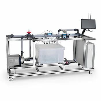
- Orifice and Venturi Flowmeter Calibration Educational Pilot Plant for Fluid Mechanics Laboratory
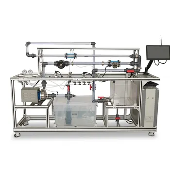
- Centrifugal Pump Performance and Orifice Flowmeter Calibration Educational Pilot Plant
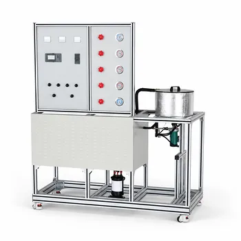
- Educational Compression Refrigeration Performance Determination Unit Operations Pilot Plant
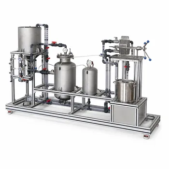
- Constant Pressure Filtration Educational Unit Operations Pilot Plant
- Fluid Friction Resistance Determination Educational Unit Operations Pilot Plant
People Also Ask
- What is the difference between static and stagnation pressure? Master Pilot Plant Flow Measurement
- How do pilot plants demonstrate siphon pressure variations? Visualizing Bernoulli's Energy Balance
- How do fluid mechanics training pilot plants facilitate the visualization and calculation of laminar and turbulent flows?
- How to update chemometric calibration models in pilot plants? Best practices for process engineers.
- Why are the laws of similitude critical in fluid flow pilot plants? Scale Up Safely