For a robust and reproducible teaching lab procedure, immobilize laccase and glucose dehydrogenase (GDH) in a polyvinyl alcohol (PVA) hydrogel via photo-polymerization. The membrane is cast from a cooled PVA solution mixed with enzyme concentrates, cured under UV light on a plastic substrate, and then mounted on an oxygen electrode behind a dialysis membrane. This protocol consistently yields functional bienzyme membranes that can demonstrate glucose-dependent oxygen consumption in stirred-cell or flow-injection biosensor setups.
The core takeaway: A simple, UV-initiated PVA immobilization—mixing enzymes into a pre‑dissolved PVA matrix, casting onto disks, and curing for ~30 minutes—creates a durable, education‑ready bienzyme membrane that couples laccase’s oxygen reduction to GDH’s glucose oxidation, making biosensor principles tangible without expensive equipment or hazardous chemicals.
The PVA Photo-Polymerization Protocol
The reference method uses a physical entrapment strategy inside a PVA network that is crosslinked by UV light. This one‑pot process keeps the workflow short enough for a single lab session.
Preparing the PVA Matrix
Start by suspending 400 mg of PVA in 2 ml of deionized water. Heat this suspension to 100 °C while stirring until the PVA dissolves completely into a clear or slightly hazy solution. Then allow the solution to cool to room temperature—this step prevents thermal denaturation of the enzymes when they are added later.
Enzyme Incorporation and Membrane Casting
Take the cooled PVA solution and mix it with equal volumes of high‑activity enzyme solutions. The reference suggests enzymes in the range of 9000 U/ml for laccase and for GDH. Gently combine to avoid forming air bubbles.
Apply a measured drop of this enzyme‑PVA blend onto a clean small plastic disk—the substrate that will support the thin membrane. The disk size should match the tip of your oxygen electrode.
Photo-Polymerization and Electrode Assembly
Expose the coated disk to UV light for approximately 30 minutes. The UV irradiation triggers the PVA chains to form a crosslinked hydrogel that physically traps the enzymes. After polymerization, peel the resulting bienzyme membrane off the disk (or leave it attached if your electrode design permits) and place it directly onto the sensing surface of an oxygen electrode.
Cover the membrane with a dialysis membrane (cut slightly larger than the electrode tip) to prevent fouling and enzyme wash‑out, then secure everything with an O‑ring. The completed biosensor can now be placed in a stirred cell or integrated into a flow cell for amperometric measurement.
Why This Protocol Suits Educational Settings
Beyond the chemistry, the protocol’s logistics align perfectly with teaching lab constraints.
Minimal Equipment, Maximum Visibility
Heating a vial, mixing solutions, and curing under a simple UV lamp are operations that students can perform safely with standard lab glassware. The visible gelation of the PVA and the clear assembly onto an electrode make each step easy to monitor and troubleshoot.
Robust, Reusable Membranes
Entrapment in PVA creates a physically stable membrane that withstands gentle handling and repeated use. In a teaching lab, where students may need multiple attempts, the membranes can be stored hydrated in buffer at 4 °C for short‑term reuse.
Understanding the Trade‑offs and Common Pitfalls
No immobilization method is flawless. Being aware of the limitations helps turn failed attempts into teachable moments.
Diffusion Barriers and Response Time
The PVA hydrogel introduces a mass‑transfer resistance. Substrates (glucose, oxygen) and products must diffuse through the gel to reach the enzymes and the electrode. This can slow the sensor’s response and reduce the apparent activity. For educational demonstrations, a slower signal actually aids observation, but if students compare turnover rates to free‑enzyme assays, they will see a discrepancy.
Enzyme Leaching Over Time
Physical entrapment relies on pore‑size exclusion. Over hours of continuous operation or under vigorous stirring, smaller enzyme molecules may slowly leach out, causing a gradual loss of signal. The dialysis membrane partially compensates, but long‑term stability is limited.
UV Exposure and Enzyme Viability
The 30‑minute UV curing is essential for PVA crosslinking, yet prolonged UV can partially inactivate enzymes through radical damage or direct photolysis. The protocol’s short irradiation time and the protective effect of the PVA solution help, but slight activity loss is inevitable. For critical quantitative work, run a control without UV (if gelation still occurs) to estimate the damage, though the reference does not detail such a step.
How to Adapt This Protocol for Your Teaching Lab
Your exact implementation should reflect your learning objectives and equipment availability. Below are goal‑oriented recommendations.
- If your primary focus is on conceptual understanding of biosensor transduction: Simplify further by pre‑casting membranes and providing pre‑assembled electrodes. Students can then focus on recording calibration curves and analyzing the cascade reaction that links glucose depletion to oxygen consumption.
- If your primary focus is on hands‑on biochemical engineering: Let students prepare the PVA matrix, explore different enzyme ratios (e.g., laccase:GDH), and test the effect of UV exposure time on signal magnitude. This turns the protocol into an inquiry‑based investigation of immobilization efficiency.
- If your primary focus is on demonstrating a complete flow‑injection analysis (FIA) system: Use the same membrane assembly but plumb the electrode into a simple FIA loop. Students can inject glucose samples and observe the peak amperometric response, directly connecting biosensor hardware to modern analytical instrumentation.
By matching the complexity to the educational goal, this PVA‑based bienzyme protocol reliably transforms an abstract biosensor concept into a concrete, measurable result.
Summary Table:
| Step | Key Parameter / Action | Purpose |
|---|---|---|
| 1. PVA Prep | 400 mg PVA in 2 ml H₂O, heat to 100°C | Dissolves polymer; cool before adding enzymes |
| 2. Enzyme Mix | 1:1 ratio of Laccase & GDH (~9000 U/ml) | Blends enzymes gently without bubbling |
| 3. Curing | 30 minutes UV light exposure | Photo-polymerizes PVA to trap enzymes |
| 4. Assembly | Cover with dialysis membrane, secure with O-ring | Prevents enzyme wash-out & electrode fouling |
Elevate Your Practical Training with LABPARK
To help your students master critical concepts in bioprocess engineering and biosensor unit operations, having the right hands-on equipment is essential.
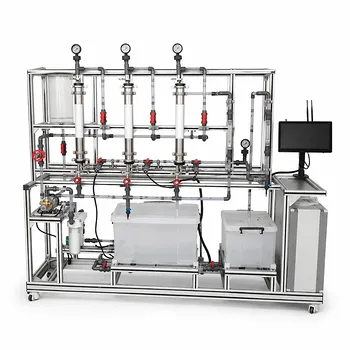
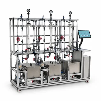
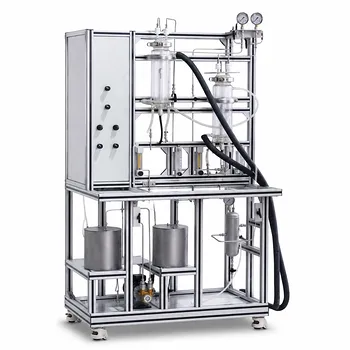
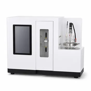
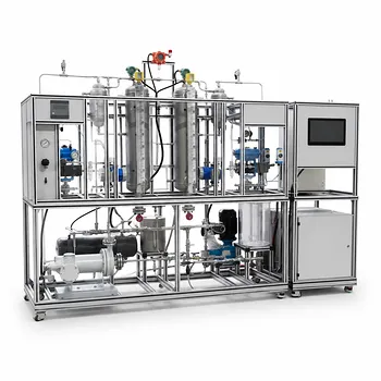
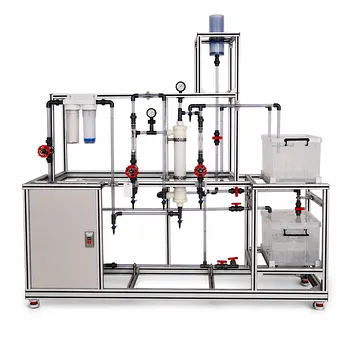
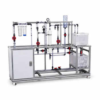
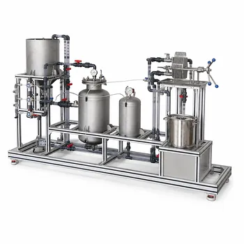
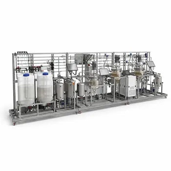
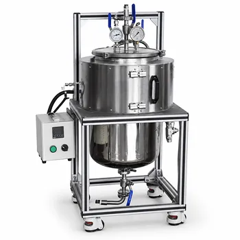
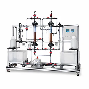
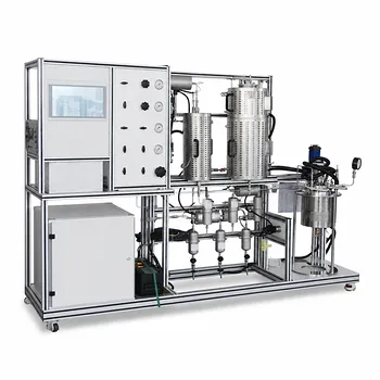
LABPARK designs and provides high-quality Educational and Vocational Unit Operations Pilot Plants in chemical engineering, bioprocess & biotech, and environmental & water treatment. Tailored for universities, research institutes, and enterprises, our systems make abstract concepts like mass-transfer limits, enzymatic cascades, and sensor calibration completely tangible.
Ready to upgrade your laboratory setup? Contact LABPARK today to discuss your curriculum needs with our technical team!
Related Products
- Multifunctional Membrane Separation Educational Pilot Plant with Ultrafiltration, Nanofiltration, Reverse Osmosis
- Multi-Functional Membrane Separation Educational Pilot Plant for Unit Operations Lab
- Photocatalytic Membrane Separation and Degradation Unit Operations Pilot Plant
- Multi Functional Membrane Crystallization Educational Unit Operations Pilot Plant
- Alkaline Membrane Water Electrolysis Educational Pilot Plant Unit Operations Training System
People Also Ask
- Pervaporation vs. Vapor Permeation: Handling Suspended Solids in Membrane Pilot Plants
- How do PEI, PVDF, and PSU membranes compare in pilot plants? Find the best fit.
- What are the trade-offs of polymer vs inorganic membranes in teaching pilot plants? Key laboratory comparison.
- How can operators optimize operating conditions and cleaning protocols in membrane separation pilot plants to mitigate fouling?
- What are the advantages of membrane separation in pilot plants? Efficient Organic Vapor Recovery