The gap in resolution between optical microscopy and transmission electron microscopy (TEM) is not just a minor technical detail—it’s a fundamental shift in what you can see. TEM can resolve features down to ~0.1 nm, while the best optical microscopes are limited by the wavelength of visible light to about 200 nm. For chemical engineering unit operations, this means the nanometer-scale metal crystallites that dictate catalyst activity, selectivity, and deactivation are completely invisible under a light microscope, whereas TEM delivers the structural detail required to engineer and troubleshoot these systems.
For analyzing active catalyst morphology, TEM's resolution eclipses optical microscopy by a factor of over 2,000, making it the only practical technique for imaging the supported metal nanoparticles that drive chemical reactions. This leap in resolving power comes with strict sample preparation requirements, contrast challenges, and the need for statistical rigor—but it is indispensable for linking catalyst structure to reactor performance.
The Physics of Resolution: Why Wavelength Determines Everything
The Fundamental Limit
The minimum distinguishable size (x) in any microscope is governed by the wavelength of the imaging beam and the spherical aberration of the lenses. For TEM, the relationship is described by:
x = 0.6 × Cs^(1/4) × λ^(3/4)
Here, λ is the wavelength of the electron beam and Cs is the spherical aberration coefficient of the electromagnetic lenses.
The Wavelength Advantage of Electrons
The power of TEM lies in the absurdly short wavelength of accelerated electrons. An electron beam has a wavelength of 0.5 to 10 Å (0.05–1 nm), while visible light spans 4000 to 7000 Å (400–700 nm).
That’s a three-orders-of-magnitude difference. An optical photon physically cannot probe features smaller than about half its wavelength—so the 2–10 nm platinum particles on a silica support remain a blur. Electrons, by contrast, easily resolve individual atomic columns.
Why This Matters for Catalyst Morphology in Chemical Engineering
The Size Regime of Active Catalysts
Supported metal catalysts in pilot plants and industrial reactors—like platinum on silica (Pt/SiO₂)—typically consist of crystallites well below 1000 Å (100 nm).
The metal particles that actually perform the catalytic chemistry often measure only 1–20 nm. Optical microscopy simply cannot detect them, let alone measure their shape or distribution.
How Morphology Links to Reactor Performance
Knowing the size distribution, particle shape, and dispersion of the active phase is critical for:
- Turnover frequency calculations that determine reaction kinetics.
- Sintering studies that explain catalyst deactivation over time.
- Structure-sensitivity analysis that guides the design of more selective catalysts.
TEM’s resolution enables you to see all of this at the atomic scale. Without it, you’d be flying blind.
Overcoming Contrast Challenges: Dark-Field Imaging in TEM
The Amorphous Support Problem
On a typical TEM grid, the thin amorphous silica or alumina support scatters electrons almost uniformly, creating very low contrast between the support and the small metal crystallites.
That’s why bright-field TEM often fails to reveal the critical particles you need to analyze.
Dark-Field Imaging as a Solution
Dark-field TEM exploits the fact that electrons diffract preferentially from the crystalline metal particles.
By using the objective aperture to select only the diffracted beams, the metal crystallites appear as bright spots on a dark, weakly diffracting support background. This transforms nearly invisible nanoparticles into countable, measurable bright features—allowing reliable size and morphology assessment even for particles barely 1 nm across.
From Dead Samples to Live Reactions: Controlled Atmosphere TEM
Ex-Situ TEM’s Fundamental Constraint
Standard TEM requires a high vacuum environment, so it can only examine catalyst samples before or after reaction.
While post-mortem analysis can detect sintering or coking, it cannot capture the real-time dynamics that cause deactivation. The catalyst structure you see under vacuum may not represent the structure that existed at 400°C under syngas pressure.
Controlled Atmosphere TEM (CATEM) Changes the Game
CATEM integrates a gas reaction cell that allows the specimen to be observed in-situ under reactive gases at pressures of several torr and temperatures up to 1300°C.
This capability enables real-time visualization of gas-solid interactions—metal particle migration, carbon filament growth, and oxidation/reduction cycles—that occur inside an actual catalytic reactor. For chemical engineers troubleshooting a pilot plant, CATEM provides the mechanistic insight that static images never could.
Understanding the Trade-offs: Where TEM Falls Short
The Statistical Sampling Challenge
A TEM image shows only an extremely small, localized region of the sample.
To obtain a statistically meaningful particle size distribution, you must analyze many micrographs—often dozens—and measure hundreds or thousands of particles. Small, low-contrast crystallites may still remain undetected under standard conditions, biasing the result toward larger particles.
The 2D Projection Trap
TEM produces two-dimensional projections of three-dimensional structures. Overlapping particles, tilted crystallites, or agglomerates can lead to misinterpretation of size and shape, especially when automated image analysis is applied without careful tilting or tomography experiments.
Sample Preparation Artifacts
Preparing catalyst powders for TEM is not trivial. The specimen must be ground to an ultrafine powder (e.g., 200 mesh) and dispersed in a liquid carrier.
Agglomeration during drying can alter the appearance of the very morphology you intend to measure, and the thin regions required for electron transparency may not be representative of the bulk catalyst.
Vacuum Incompatibility
As already noted, conventional TEM is blind to the working state of the catalyst. Only CATEM or advanced environmental TEM techniques bridge this gap, and they introduce complexity in temperature control, gas purity, and image interpretation.
Making the Right Choice for Your Catalyst Analysis Goal
Choose your approach based on the specific engineering question you need to answer.
- If your primary focus is resolving nanoparticle size, shape, and lattice structure at the atomic level: Conventional TEM with dark-field imaging is the undisputed standard. Pair it with careful statistical analysis of many micrographs to ensure representativeness.
- If your primary focus is observing catalyst behavior under reaction conditions (sintering, coking, metal–support interactions): Invest in Controlled Atmosphere TEM or seek access to a facility with in-situ capabilities. The real-time mechanistic data can transform your understanding of deactivation pathways.
- If your primary focus is obtaining a statistically robust, bulk-averaged size distribution: Remember that TEM alone is insufficient. Combine TEM-derived particle counts with bulk techniques like chemisorption or X-ray diffraction, and use multiple grid preparations to minimize sampling bias.
TEM doesn’t just see smaller—it reveals the hidden architecture that governs catalytic performance, but only a strategic, statistically grounded approach turns its extreme resolution into actionable engineering insight.
Summary Table:
| Feature | Optical Microscopy | Transmission Electron Microscopy (TEM) |
|---|---|---|
| Resolution Limit | ~200 nm | ~0.1 nm |
| Imaging Source | Visible light (400-700 nm) | Electron beam (0.05-1 nm) |
| Catalyst Visibility | Invisible (<100 nm particles) | Highly visible (down to 1 nm) |
| Environment | Ambient / liquid | High vacuum (CATEM for controlled gas) |
Optimize Your Unit Operations with LABPARK
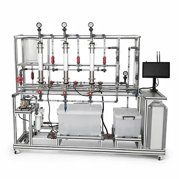
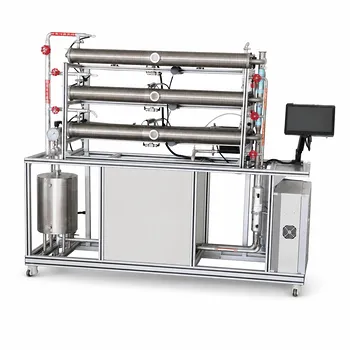
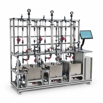
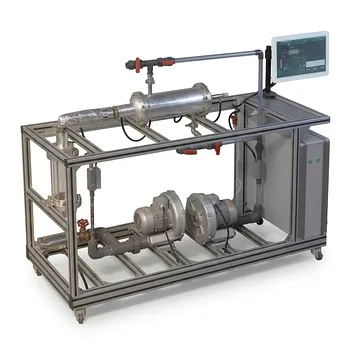
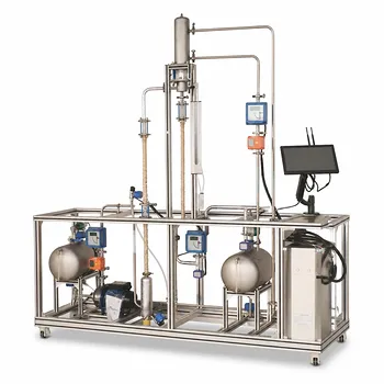
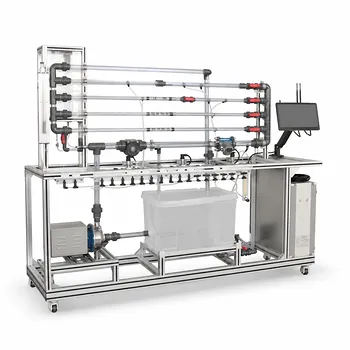
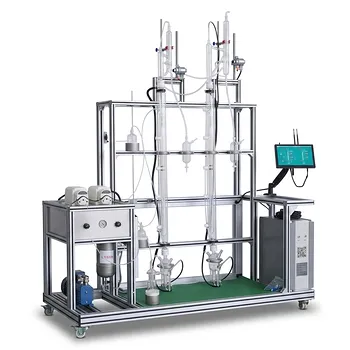
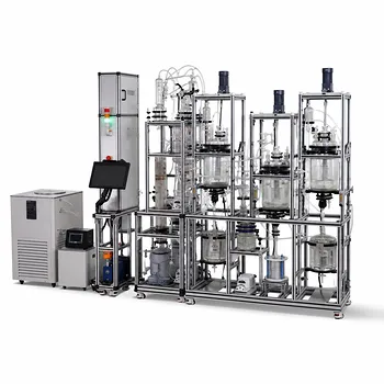
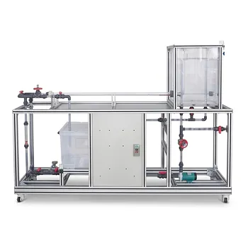
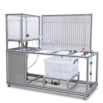
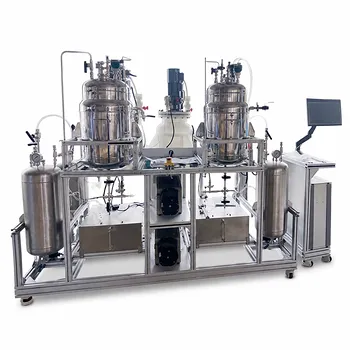
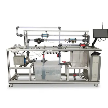
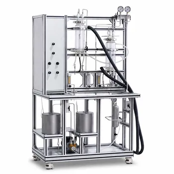
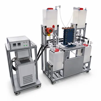
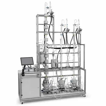
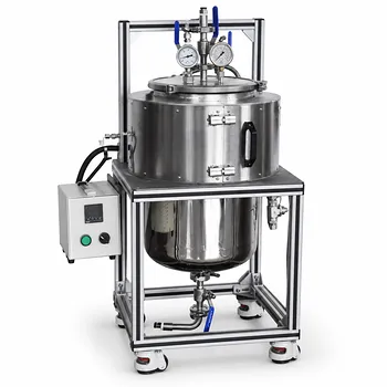
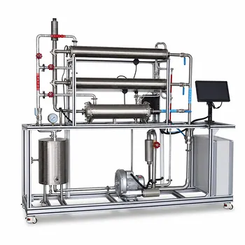
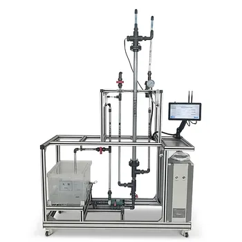
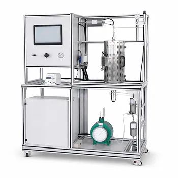
Bridging the gap between atomic-scale catalyst research and industrial-scale unit operations requires the right hands-on training and testing systems. LABPARK provides state-of-the-art Educational and Vocational Unit Operations Pilot Plants in chemical engineering, bioprocess & biotech, and environmental & water treatment.
Designed specifically for universities, research institutes, and enterprises, our pilot plants help you translate nanoscale insights into optimized, real-world processes.
Ready to elevate your research and engineering training? Contact us today to discover our custom pilot plant solutions!
Related Products
- Multifunctional Membrane Separation Educational Pilot Plant with Ultrafiltration, Nanofiltration, Reverse Osmosis
- Three-Tube Heat Transfer Educational Pilot Plant for Unit Operations Training
- Multi-Functional Membrane Separation Educational Pilot Plant for Unit Operations Lab
- Shell and Tube Heat Exchanger Heat Transfer Coefficient Determination Educational Pilot Plant
- Rising and Falling Film Evaporation Educational Unit Operations Pilot Plant
People Also Ask
- Pervaporation vs. Vapor Permeation: Handling Suspended Solids in Membrane Pilot Plants
- How do PEI, PVDF, and PSU membranes compare in pilot plants? Find the best fit.
- What are the trade-offs of polymer vs inorganic membranes in teaching pilot plants? Key laboratory comparison.
- What are the key flow configuration loops in membrane pilot plants? Optimize Performance
- How can operators optimize operating conditions and cleaning protocols in membrane separation pilot plants to mitigate fouling?