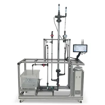
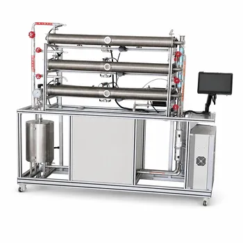
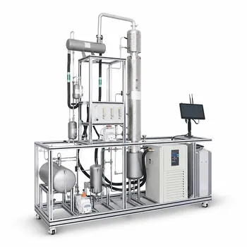
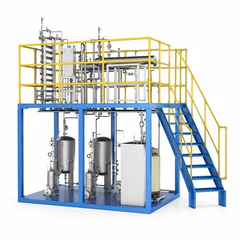
Electron wave-particle duality isn’t just a physics puzzle—it’s the operational principle that equips unit operations pilot plants with the ability to see the nanoscale reality that governs their performance. In university biotechnology and chemical engineering labs, this concept directly underpins the electron microscopes that students use to inspect membrane fouling, catalyst surfaces, and cell morphologies. By exploiting the wave nature of accelerated electrons, these analytical modules resolve features a thousand times smaller than the diffraction limit of visible light, turning abstract quantum behavior into a concrete engineering tool.
The de Broglie wavelength of an electron transforms a particle into a probe whose resolution can be tuned with voltage. In a pilot plant, this means a scanning electron microscope (SEM) can image the root cause of a pressure drop, a transmission electron microscope (TEM) can visualize catalyst deactivation at the atomic scale, and image‑based particle sizing can reveal aggregation states—bridging foundational quantum mechanics and day‑to‑day process troubleshooting.
Why Traditional Optics Hit a Wall in Pilot Plants
The Resolution Barrier of Visible Light
Conventional optical microscopes are limited by diffraction to a resolution of roughly 200 nanometers. Many critical features in unit operations—pores in an ultrafiltration membrane, surface asperities on a catalyst pellet, or microvilli on a cell—are far smaller. To see them, you need a source with a much shorter effective wavelength.
de Broglie’s Equation: Turning Mass into Magnification
An electron accelerated through a potential difference gains kinetic energy, and its momentum defines a wavelength given by λ = h/p. The de Broglie wavelength of a 10 kV electron is already around 0.012 nm—three orders of magnitude smaller than visible light. This is the core of electron wave‑particle duality in the lab: the same entity that behaves as a particle in a circuit becomes a wave that can be focused into an imaging beam, with resolution ultimately limited only by the optics and the sample.
The Analytical Toolkit Powered by Electron Duality
Scanning Electron Microscopy (SEM): The Workhorse of Unit Ops
SEM scans a finely focused electron beam over a sample and collects signals such as secondary electrons and backscattered electrons. This yields topographical and compositional images down to the nanoscale. In a pilot plant setting, students routinely use SEM to examine:
- Membrane cross‑sections: revealing fouling layer thickness, pore blockage, and gel‑layer structure.
- Catalyst particles: showing particle size, morphology, and dispersion on a support.
- Bioreactor surfaces: visualizing cell attachment and extracellular matrix on microcarriers or hollow fibers.
Transmission Electron Microscopy (TEM) for Internal Structures
For even finer detail, TEM sends electrons through an ultra‑thin specimen. Its wave properties enable atomic‑scale resolution, making it indispensable for:
- Catalyst characterization: imaging metal nanoparticle size distributions and identifying sintering or carbon encapsulation.
- Biotechnology: examining intracellular organization or viral vector morphology. Because electron wavelength decreases with accelerating voltage, a 200 kV beam brings individual atomic columns into clear view.
Electron‑Beam‑Based Particle Sizing (via Image Analysis)
While dynamic light scattering is a bulk method, electron microscopy provides direct measurement of particle size, shape, and aggregation from images. By applying automated image‑analysis software to SEM or TEM micrographs, students obtain number‑based size distributions with visual context that light‑based techniques cannot offer. This is the “advanced particle sizing” capability enabled by the wave nature of electrons—direct morphological evidence, not just a statistical average.
Seeing the Invisible: Practical Applications in Unit Operations
Membrane Filtration: Diagnosing Fouling at the Root
When pilot plant flux declines unexpectedly, a pressure sensor tells you something is wrong, but not what. An SEM cross‑section reveals the culprit: a dense calcium sulfate scaling layer, a biofilm, or a compressible gel. Knowing the fouling mechanism allows students to select an appropriate cleaning protocol or modify membrane chemistry—decisions grounded in tangible, wave‑enabled visualization.
Heterogeneous Catalysis: Tracking Deactivation
A packed‑bed reactor’s conversion might drop, but bulk assays only hint at the cause. TEM images of spent catalyst can directly show particle sintering, carbon whisker growth, or metal leaching. These nanoscale observations link the performance loss to a physical change, guiding regeneration procedures or prompting a redesign of the catalyst support.
Bioreactors: Cell Morphology and Surface Interactions
In a perfusion bioreactor, healthy cells must attach and spread properly. SEM images of harvested microcarriers expose whether cells have formed a confluent monolayer or whether apoptosis‑related blebbing is present. Because electron waves penetrate only a few nanometers into the surface, the image provides exceptional surface sensitivity—exactly what’s needed to assess cell‑material interactions.
Understanding the Trade‑offs of Electron‑Based Analysis
The Vacuum Requirement and Sample Preparation
Electron beams cannot travel far in air, so samples must be placed in a high vacuum. For biological or hydrated specimens, this means chemical fixation, dehydration, and metal coating. These steps can introduce artifacts, such as membrane pore collapse or cell shrinkage. Cryo‑SEM and ESEM offer workarounds, but they trade some resolution for the ability to image partially hydrated samples.
Cost, Footprint, and Expertise
High‑resolution SEM and TEM instruments are capital‑intensive, require dedicated space and utilities, and demand skilled operators. In a teaching pilot plant, this means electron microscopy is typically an offline module where extracted samples are analyzed in a separate facility, not a real‑time inline sensor.
Potential Beam Damage
Polymers, proteins, and delicate biological structures can be destroyed by the electron beam’s energy. Students must work at low beam currents or apply dose‑minimization strategies, accepting that the sharpest images sometimes come at the cost of a slightly altered specimen.
Statistical Sampling vs. Bulk Methods
An SEM image covers a minuscule area. A single micrograph may not represent the entire membrane surface or catalyst batch. Pairing electron microscopy with bulk techniques—laser diffraction for particle size, chemisorption for active site density, or TOC for fouling mass—is essential to validate that the viewed area is typical and to build a statistically robust picture.
Making Analytical Modules Work for Your Pilot Plant Goal
How you deploy these wave‑enabled analytical modules depends on what you need to solve. Use this decision guide to align the tool with the problem.
- If your primary focus is troubleshooting membrane performance: Use SEM cross‑sectional imaging to directly measure fouling layer thickness and morphology. Combine with real‑time pressure‑drop data to correlate structural evolution with hydraulic resistance.
- If your primary focus is catalyst development or lifetime studies: Plan regular TEM sampling to track nanoparticle size and distribution. Overlay this with activity and selectivity data to identify the deactivation mechanism, whether it be sintering, coking, or poisoning.
- If your primary focus is bioprocess optimization: Choose cryo‑fixation or low‑vacuum SEM to preserve cell and matrix structure. Use the resulting images to validate CFD models of shear stress around microcarriers or to confirm the integrity of a 3D‑printed scaffold.
- If your primary focus is particle size distribution for quality control: Use SEM‑image analysis to understand shape and aggregation, but validate with a bulk method like dynamic light scattering or laser diffraction to ensure the sampled area represents the whole batch.
By mastering the analytical modules born from electron wave‑particle duality, you transform pilot plant run data into a visual narrative of nanoscale events—enabling the kind of informed, root‑cause‑driven design that textbooks alone cannot teach.
Summary Table:
| Analytical Module | Duality Application | Main Pilot Plant Use | Key Limitation |
|---|---|---|---|
| SEM | Surface scanning with focused electron waves | Membrane fouling, catalyst morphology, cell attachment | High vacuum required, surface-only |
| TEM | Electron transmission through thin samples | Catalyst deactivation (sintering), intracellular structures | Extensive sample prep, potential beam damage |
| Image-Based Sizing | Direct imaging & software-based analysis | Number-based particle size & shape distribution | Localized sampling (lacks bulk statistical weight) |
Elevate Your Lab's Analytical & Process Capabilities with LABPARK
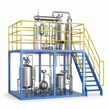
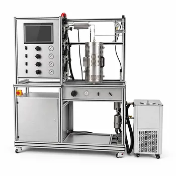
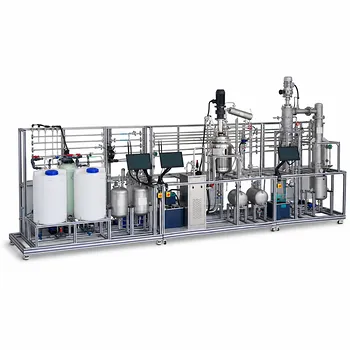
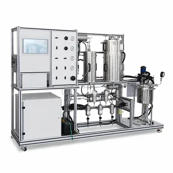
Bridging quantum theory and practical engineering requires high-performance equipment. LABPARK provides state-of-the-art Educational and Vocational Unit Operations Pilot Plants in chemical engineering, bioprocess & biotech, and environmental & water treatment.
Designed specifically for universities, research institutes, and enterprises, our pilot plants seamlessly integrate advanced process control with analytical modules to help your students and researchers visualize nanoscale phenomena and optimize real-world processes.
Ready to upgrade your laboratory training and research infrastructure? Contact us today to explore our custom pilot plant solutions!
Related Products
- Multi-Modal Distillation Unit Operations Training Pilot Plant
- Green Anhydrous Ethanol Purification Extractive Distillation Unit Operations Training Pilot Plant
- Comprehensive Multi-Modal Heat Transfer Unit Operations Pilot Plant for Engineering Training
- Ethyl Acetate Synthesis Unit Operations Pilot Plant for Practical Training
- Dual-Mode Rectification Pilot Plant for Practical Training Unit Operations
People Also Ask
- How can energy balance equations for condensers & reboilers be validated experimentally? Guide
- How do distillation pilot plants validate VLE data? Achieve reliable solvent separation scale-up.
- How can real-time carbon number prediction improve distillation pilot plants? Optimize control.
- Why is the column performance diagram critical? Optimize distillation pilot plant safety & efficiency.
- What are the primary reflux ratio control strategies? Master Distillation Unit Operations