Identifying a single target protein in a complex bioprocess mixture can feel like finding a needle in a haystack. A dual-detector setup that pairs a standard UV absorbance monitor with an acoustic wave sensor fundamentally solves this problem. The acoustic sensor generates a cumulative phase signal that rises only when your protein of interest binds to its surface. By taking the first derivative of that integral signal, you create a peak-based profile that aligns with elution times, and overlaying it on the conventional UV chromatogram instantly reveals which UV peak corresponds to your target—no labels, no guesswork.
When UV traces become an unreadable tangle of overlapping peaks, an acoustic wave sensor brings molecular specificity. It accumulates a binding signal exclusively from your target. Transforming that signal into a derivative curve and superimposing it on the UV chromatogram gives you a clear, label-free method to pinpoint the exact peak you’re after, even when it co-elutes with background proteins.
The Core Challenge: UV Blindness in Complex Mixtures
In downstream processing, your chromatography run often deals with raw or partially purified streams—think proteins from fetal bovine serum or cell culture harvests. A UV detector measures total absorbance, so every protein that passes through the flow cell contributes to the signal. When dozens of species elute at similar times, the resulting chromatogram becomes a series of overlapping peaks where your target is invisible.
Why a Single UV Trace Tells You Almost Nothing
The UV signal is not specific. It reflects the combined absorbance of all co-eluting molecules.
A shoulder on a major peak, a slight hump in the baseline, or a cluster of fused peaks can all contain your protein, but you have no way to identify which one is yours without additional assays.
This blindness forces you to collect fractions and run time-consuming offline tests, slowing development and introducing risk.
The Deep Need: Real‑Time, Label‑Free Identification
Operators don’t just need a chromatogram; they need to know with certainty when their target elutes.
This demands a detection method that responds only to the analyte of interest, works in real time, and does not require fluorescent or enzymatic labels.
That is exactly where a dual‑detector strategy with an acoustic wave sensor comes in.
How the Acoustic Wave Sensor Provides Target Specificity
An acoustic wave sensor—often based on a quartz crystal microbalance or surface acoustic wave technology—measures mass changes on its surface by tracking shifts in resonant frequency or phase. When the sensor is functionalized with a ligand (an antibody, aptamer, or other capture molecule) that selectively binds your target protein, it becomes a target‑specific detector.
The Cumulative Phase Signal: A Direct Measure of Binding
As your protein of interest binds to the sensor surface, the increasing mass produces a monotonically rising phase signal.
This is an integral or cumulative response: it keeps growing as long as more target attaches.
Crucially, because the sensor only captures the target protein, the signal is completely blind to all other components in the mixture. You get a clean, specific trace that represents nothing but your analyte.
Derivative Transformation: From Integral to Peak‑Based Elution Profiles
A cumulative curve is hard to align with a traditional chromatogram, which displays peaks.
The solution is simple: take the first derivative of the cumulative phase signal with respect to time.
The derivative shows the rate of mass addition. This rate spikes when a concentrated band of target protein flows over the sensor—exactly at its elution time—and falls back to zero when the band passes. The result is a peak‑shaped profile that mirrors the elution pattern of the target.
Overlaying with UV: Pinpointing the Peak of Interest
Now you have two time‑aligned signals: the messy UV trace with multiple overlapping peaks, and the sharp derivative curve that peaks only where your target elutes.
By superimposing them on the same time axis, the derivative signal acts like a highlighting marker.
The UV peak that coincides with the derivative maximum is your target protein. Even if it hides as a small shoulder on a much larger peak, the overlay eliminates ambiguity.
Understanding the Trade‑offs and Practical Considerations
While powerful, this dual‑detector approach is not a magic bullet. Being aware of its boundaries ensures you apply it successfully.
Sensor Surface Regeneration and Lifetime
The binding surface must be regenerated between runs without damaging the capture molecule.
Harsh regeneration conditions can degrade performance over time, requiring careful buffer selection and validation.
Potential Baseline Drift and Noise
A cumulative signal inherently amplifies any slow, non‑specific adsorption or temperature effects over a long run.
This can distort the derivative curve unless corrected. Implementing a reference sensor or signal processing steps is often necessary to maintain reliable peak identification.
Ligand Specificity and Availability
The technique relies entirely on having a ligand that binds your target selectively and with sufficient affinity.
If such a ligand does not exist or cross‑reacts with matrix components, the sensor loses its specificity, and the method collapses back to a non‑informative signal.
Complexity in Multiplexing
For more than one target, you would need multiple sensor channels or sequential assays.
While feasible, this adds instrument complexity and cost compared to a single UV detector.
Making the Right Choice for Your Bioprocess Goal
How you implement a dual‑detector setup depends on what you aim to achieve. The following recommendations help you focus your efforts.
- If your primary focus is rapid, label‑free identification of a known target in a dirty sample: Prioritize an acoustic wave sensor functionalized with a robust, high‑affinity capture molecule. The derivative‑overlay method will immediately tell you which fraction to pool or which peak to integrate for quantification, without any offline ELISA‑style testing.
- If your primary focus is quantitative purity analysis alongside identification: Combine the dual‑detector data with a calibration curve derived from the acoustic sensor’s maximum binding rate (the derivative peak height). This enables concentration estimation directly from the specific signal, turning the setup into a combined identity and quantity check.
- If your primary focus is early‑stage process development with limited time and resources: Use the dual‑detector approach to drastically cut down on fraction collection and analysis. By confirming target elution in real time, you can iterate chromatography conditions faster and with higher confidence, even when the UV chromatogram shows no clear resolution.
A single UV detector leaves you guessing; an acoustic wave sensor gives you eyes that see only what matters. Used together, they transform an overlapping mess into a clear, actionable map of your target protein’s journey.
Summary Table:
| Feature | UV Detector | Acoustic Wave Sensor | Dual-Detector Setup |
|---|---|---|---|
| Specificity | Low (measures total absorbance) | High (target-specific binding) | High (pinpoints target in complex mixtures) |
| Signal Output | Peak-based (chromatogram) | Cumulative (integral phase signal) | Peak-based (via first derivative) |
| Elution Tracking | Blind to co-eluting impurities | Tracked, but lacks chromatographic context | Instantly aligns target peak with UV trace |
Scale Up Your Bioprocess Training and Research with LABPARK
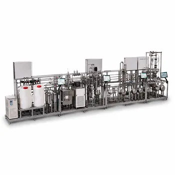
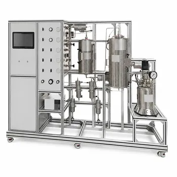
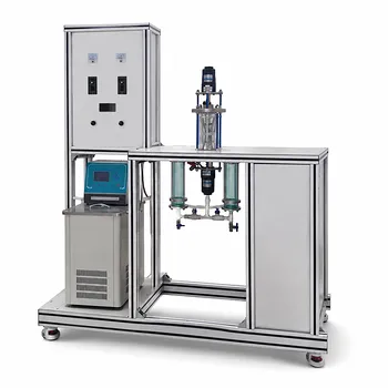
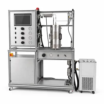
Developing expertise in advanced protein purification and detection requires hands-on experience with industry-grade equipment. LABPARK provides premium Educational and Vocational Unit Operations Pilot Plants in chemical engineering, bioprocess & biotech, and environmental & water treatment.
Designed specifically for universities, research institutes, and enterprises, our pilot plants empower students and researchers to master complex separation, filtration, and analytical workflows.
Ready to elevate your lab's capabilities? Contact LABPARK today to find the perfect pilot plant solution for your institution.
Related Products
- Electrolyte Distillation Purification and Formulation Educational Pilot Plant
- Multifunctional Membrane Separation Educational Pilot Plant with Ultrafiltration, Nanofiltration, Reverse Osmosis
- Multi-Functional Membrane Separation Educational Pilot Plant for Unit Operations Lab
- Multi Functional Catalytic Reaction and Reactor Evaluation Educational Unit Operations Pilot Plant
- 100L Continuous Loop Hydrogenation Educational Unit Operations Pilot Plant
People Also Ask
- Why does simple distillation yield higher efficiency than flash distillation? Key thermodynamic differences.
- How to Integrate Spectroscopy in Distillation Pilot Plants for Advanced Process Control
- How does double temperature difference control improve distillation pilot plants? Stabilize purity.
- How does the feed thermal state influence distillation pilot plant design and utility consumption?
- How to Estimate Distillation Pilot Plant Tray Efficiency? O'Connell's Correlation Guide